
Home » Nervous system

Custom Search
Popular Posts
-
 Bursae act as friction-reducing cushions around joints. Inflammation of a bursa, called bursitis, may occur if it is put under prolonged or ...
-
 Infection of the testis and epididymis (the coiled tube that stores and transports sperm away from the testis) can lead to inflammation call...
-
 If large areas of both sides of the brain or the brainstem are damaged, coma may result. In persistent vegetative state, the parts of the br...
-
Every women differs and so differs there choice of foreplay, some love it to be soft and tender while others relish wild foreplay. Kissing i...
-
 A ganglion is a cyst that develops under the skin near a joint and is filled with a jelly-like fluid. The ganglion is usually an outgrowth f...
-
 Normally, vaginal bleeding occurs only during a period. Bleeding that occurs outside of menstruation is abnormal. In women under the age o...
-
 Wernicke-Korsakoff syndrome is a rare disorder of the brain, causing dementia, abnormal eye movements, and an abnormal gait. The condition d...


Facial palsy
The facial nerve controls the muscles of expression and emotion in the face and caries taste sensations from the front of the tongue to the brain. In facial palsy, one of the two facial nerves is damaged, compressed, or inflamed, and this results in weakness of the facial muscles, causing the eyelid and corner of the mouth to droop on one side of the face. People with facial palsy are often concerned that they have had a stroke, but this is unlikely if only the face is affected because a stroke is usually also associated with muscle weakness in other parts of the body.

Facial palsy is usually temporary, but a full recovery may take several months. The disorder affects about 1 in 4,000 people in the UK each year.
What are the causes?
The most common form of facial palsy is Bell’s palsy. This type of palsy occurs for no known reason, although a viral infection is believed to play a part.
In order types of facial palsy, there are known causes of damage to the facial nerve. These include the viral infection shingles and the bacterial infection Lyme disease. In addition, the facial nerve sometimes becomes inflamed as a result of middle-ear infection. In rare cases, the facial nerve may be compressed by a tumour called an acoustic neuroma. Facial palsy can also result from damage to the nerve from a tumour of the paroid salivary gland.
What are the symptoms?
In some cases, such as in Bell’s palsy, the symptoms of facial palsy appear suddenly over about 24 hours. In other cases, including facial palsy caused by an acoustic neuroma, symptoms may develop slowly. The symptoms include:
- Partial or complete paralysis of the muscles on one side of the face.
- Pain behind the ear on the affected side of the face.
- Drooping of the corner of the mouth, sometimes associated with drooling.
- Inability to close the eyelid on the affected side and watering of the eye.
- Impairment of taste.
If facial palsy is very severe, you may have difficulty in speaking and eating, and, occasionally, sounds may seem unnaturally loud in the ear on the affected side. If the eyelid cannot be closed, the eye may become infected, leading to ulceration of the cornea, the transparent front part of the eye. In facial palsy due to shingles, you will also have a rash of crusting blisters on your ear.
How is it diagnosed?
Your doctor will probably be able to diagnose facial palsy from your symptoms alone. A rapid onset over about 24 hours suggests Bell’s palsy. Symptoms that develop more slowly usually indicate another cause.
If your doctor suspects a tumour may be compressing the facial nerve, he or she may arrange for you to have CT scanning or MRI. Nerve and muscle electrical tests may also be arranged to assess nerve damage. If you live in a part of the UK where Lyme disease is common, you may have a blood test to look for evidence of this disorder.
What might be done?
If your symptoms have appeared in the last 48 hours, your doctor may prescribe corticosteroids for up to 2 weeks to reduce inflammation of the nerve. He or she may also recommend that you take painkillers. To prevent damage to the cornea, you may be given artificial tears, and you will probably be advised to tape the affected eye shut when you go to sleep.
Bell’s palsy usually clears up without further treatment. If facial palsy has an underlying cause, it will be treated if possible. For example, if facial palsy is due to shingles, antiviral drugs, such as aciclovir, will be prescribed. To be effective, treatment with acyclovir should begin as soon as the rash appears. If you have an acoustic neuroma, it will be removed surgically to relieve compression of the facial nerve.
If muscle paralysis persists, plastic surgery may be used to reroute another nerve to the face. Facial exercises and massage may help to maintain tone.
What is the prognosis?
With appropriate treatment, facial palsy usually improves within about 2 weeks. However, a full recovery may take up to 3 months. Some people are left with weakness, and facial palsy may recur.
Read more…
Facial palsy is usually temporary, but a full recovery may take several months. The disorder affects about 1 in 4,000 people in the UK each year.
What are the causes?
The most common form of facial palsy is Bell’s palsy. This type of palsy occurs for no known reason, although a viral infection is believed to play a part.
In order types of facial palsy, there are known causes of damage to the facial nerve. These include the viral infection shingles and the bacterial infection Lyme disease. In addition, the facial nerve sometimes becomes inflamed as a result of middle-ear infection. In rare cases, the facial nerve may be compressed by a tumour called an acoustic neuroma. Facial palsy can also result from damage to the nerve from a tumour of the paroid salivary gland.
What are the symptoms?
In some cases, such as in Bell’s palsy, the symptoms of facial palsy appear suddenly over about 24 hours. In other cases, including facial palsy caused by an acoustic neuroma, symptoms may develop slowly. The symptoms include:
- Partial or complete paralysis of the muscles on one side of the face.
- Pain behind the ear on the affected side of the face.
- Drooping of the corner of the mouth, sometimes associated with drooling.
- Inability to close the eyelid on the affected side and watering of the eye.
- Impairment of taste.
If facial palsy is very severe, you may have difficulty in speaking and eating, and, occasionally, sounds may seem unnaturally loud in the ear on the affected side. If the eyelid cannot be closed, the eye may become infected, leading to ulceration of the cornea, the transparent front part of the eye. In facial palsy due to shingles, you will also have a rash of crusting blisters on your ear.
How is it diagnosed?
Your doctor will probably be able to diagnose facial palsy from your symptoms alone. A rapid onset over about 24 hours suggests Bell’s palsy. Symptoms that develop more slowly usually indicate another cause.
If your doctor suspects a tumour may be compressing the facial nerve, he or she may arrange for you to have CT scanning or MRI. Nerve and muscle electrical tests may also be arranged to assess nerve damage. If you live in a part of the UK where Lyme disease is common, you may have a blood test to look for evidence of this disorder.
What might be done?
If your symptoms have appeared in the last 48 hours, your doctor may prescribe corticosteroids for up to 2 weeks to reduce inflammation of the nerve. He or she may also recommend that you take painkillers. To prevent damage to the cornea, you may be given artificial tears, and you will probably be advised to tape the affected eye shut when you go to sleep.
Bell’s palsy usually clears up without further treatment. If facial palsy has an underlying cause, it will be treated if possible. For example, if facial palsy is due to shingles, antiviral drugs, such as aciclovir, will be prescribed. To be effective, treatment with acyclovir should begin as soon as the rash appears. If you have an acoustic neuroma, it will be removed surgically to relieve compression of the facial nerve.
If muscle paralysis persists, plastic surgery may be used to reroute another nerve to the face. Facial exercises and massage may help to maintain tone.
What is the prognosis?
With appropriate treatment, facial palsy usually improves within about 2 weeks. However, a full recovery may take up to 3 months. Some people are left with weakness, and facial palsy may recur.
Carpal tunnel syndrome
The carpal tunnel is the narrow space formed by the bones of the wrist (carpal bones) and the strong ligament that lies over them. In carpal tunnel syndrome, the median nerve, which controls some hand muscles and conveys sensation from nerve endings in part of the hand, is compressed where it passes through the tunnel. This compression causes painful tingling in the hand, wrist, and forearm. Carpal tunnel syndrome is a common disorder, especially in women aged 40-60, and often affects both hands.
What are the causes?
In some cases, the underlying cause of nerve compression is not known. In others, it occurs because the soft tissue within the carpal tunnel swell, compressing the median nerve at the wrist. Such swelling may be due to diabetes mellitus, or it may occur during pregnancy. The carpal tunnel may also be narrowed by a joint disorder, such as rheumatoid arthritis, or by a wrist fracture. The syndrome is associated with work that involves repetitive hand movements, such as typing, which can result in inflammation of the tendons in the wrist.

What are the symptoms?
Symptoms mainly affect specific areas of the hand, such as thumb, the first and middle fingers, and the palm of the hand. Initially, symptoms may include:
- Burning and tingling in the hand.
- Pain in the wrist and up the forearm.
As the condition worsens, other symptoms may gradually appear including:
- Numbness of the hand.
- Weakened grip.
- Wasting of some hand muscles, particularly at the base of the thumb.
Symptoms may be more severe at night, and pain may interrupt sleep. Shaking the affected arm may temporarily relieve symptoms, but the numbness may become persistent if left untreated.
What might be done?
Your doctor may suspect carpal tunnel syndrome from your symptoms. He or she will examine your wrists and hands may tap the inside of your wrists to check if a tingling sensation occurs. Nerve conduction studies may be carried out to confirm the diagnosis. If pregnancy is the cause of carpal tunnel syndrome, the symptoms usually disappear after childbirth. In other cases, treating the cause, if it can be identified, usually relieves symptoms.
The symptoms of carpal tunnel syndrome may be relieved temporarily by nonsteroidal anti-inflammaroty drugs or by wearing a wrist splint, particularly at night. In some cases, a corticosteroid injection under or around the ligament may reduce swelling. If symptoms persist or recur, you may have surgery under local anaesthesia to cut the ligament and release pressure on the nerve. After surgery, there are usually no further symptoms.
Read more…
What are the causes?
In some cases, the underlying cause of nerve compression is not known. In others, it occurs because the soft tissue within the carpal tunnel swell, compressing the median nerve at the wrist. Such swelling may be due to diabetes mellitus, or it may occur during pregnancy. The carpal tunnel may also be narrowed by a joint disorder, such as rheumatoid arthritis, or by a wrist fracture. The syndrome is associated with work that involves repetitive hand movements, such as typing, which can result in inflammation of the tendons in the wrist.
What are the symptoms?
Symptoms mainly affect specific areas of the hand, such as thumb, the first and middle fingers, and the palm of the hand. Initially, symptoms may include:
- Burning and tingling in the hand.
- Pain in the wrist and up the forearm.
As the condition worsens, other symptoms may gradually appear including:
- Numbness of the hand.
- Weakened grip.
- Wasting of some hand muscles, particularly at the base of the thumb.
Symptoms may be more severe at night, and pain may interrupt sleep. Shaking the affected arm may temporarily relieve symptoms, but the numbness may become persistent if left untreated.
What might be done?
Your doctor may suspect carpal tunnel syndrome from your symptoms. He or she will examine your wrists and hands may tap the inside of your wrists to check if a tingling sensation occurs. Nerve conduction studies may be carried out to confirm the diagnosis. If pregnancy is the cause of carpal tunnel syndrome, the symptoms usually disappear after childbirth. In other cases, treating the cause, if it can be identified, usually relieves symptoms.
The symptoms of carpal tunnel syndrome may be relieved temporarily by nonsteroidal anti-inflammaroty drugs or by wearing a wrist splint, particularly at night. In some cases, a corticosteroid injection under or around the ligament may reduce swelling. If symptoms persist or recur, you may have surgery under local anaesthesia to cut the ligament and release pressure on the nerve. After surgery, there are usually no further symptoms.
Sciatica
Sciatica is a form of nerve pain that may be felt anywhere along the course of one of the sciatic nerves, the two largest nerves in the body and the main nerve in each leg. The sciatic nerves are formed from nerve roots in the lower part of the spinal cord. They run from the base of the spine down the backs of the thighs to above the knees, where they divide into branches that supply the front and back of the leg and foot. The pain of sciatica is caused by compression of or damage to the sciatic nerve, usually where it leaves the spinal cord. Many people have at least one episode of sciatica during their lives. Often, only one leg is affected. In most cases, the pain disappears gradually over about 1-2 weeks, but it may recur.
What are the causes?
Sometimes, the cause of sciatica is unknown. However, in people aged 20 to 40, the most common cause is a prolapsed or herniated disc in the spinal column that presses on a spinal nerve root. Sciatica may also develop from spondylolisthesis, in which a vertebra (bone in the spine) slips out of position. In older people, sciatica may be caused by changes in the spine as a result of various conditions, such as osteoarthritis. Women may develop sciatica during the last months of pregnancy because of posture changes that cause increased pressure on the sciatic nerve.
Muscle spasm and sitting in an awkward position for long periods of time are relatively common causes of brief episodes of sciatica in all age groups.

Less commonly, sciatica may be the result of an injection into the buttock muscles that was mistakenly given too close to one of the sciatic nerves. Rarely, a tumour on the spinal cord may press on the sciatic nerve roots.
What are the symptoms?
The symptoms can be mild or severe, with spasmodic or persistent pain in the affected leg. Symptoms may include:
- Pain that is made worse by movement or by coughing.
- Tingling or numbness.
- Muscle weakness.
If sciatica is severe, you may have difficulty in lifting the foot on the affected side because of muscle weakness, and you may be unable to stand upright. Some people have difficulty in walking.
What might be done?
The doctor will examine you and test your leg reflexes, muscle strength, and sensation. You may be advised to rest in bed on a firm mattress for a couple of days and be given painkillers to relieve discomfort. If symptoms persist, you may have tests, such as MRI of the spine, to look for changes in the bones or a prolapsed disc.
Sciatica as a result of pregnancy will usually disappear after childbirth. Pain caused by muscle spasm or sitting awkwardly also tends to clear up without treatment. Occasionally, the condition can be helped by regular physiotherapy, exercise, or chiropractic treatments. However, sciatica can recur. In some cases, surgery may be necessary to relieve pressure on the nerve.
Read more…
What are the causes?
Sometimes, the cause of sciatica is unknown. However, in people aged 20 to 40, the most common cause is a prolapsed or herniated disc in the spinal column that presses on a spinal nerve root. Sciatica may also develop from spondylolisthesis, in which a vertebra (bone in the spine) slips out of position. In older people, sciatica may be caused by changes in the spine as a result of various conditions, such as osteoarthritis. Women may develop sciatica during the last months of pregnancy because of posture changes that cause increased pressure on the sciatic nerve.
Muscle spasm and sitting in an awkward position for long periods of time are relatively common causes of brief episodes of sciatica in all age groups.
Less commonly, sciatica may be the result of an injection into the buttock muscles that was mistakenly given too close to one of the sciatic nerves. Rarely, a tumour on the spinal cord may press on the sciatic nerve roots.
What are the symptoms?
The symptoms can be mild or severe, with spasmodic or persistent pain in the affected leg. Symptoms may include:
- Pain that is made worse by movement or by coughing.
- Tingling or numbness.
- Muscle weakness.
If sciatica is severe, you may have difficulty in lifting the foot on the affected side because of muscle weakness, and you may be unable to stand upright. Some people have difficulty in walking.
What might be done?
The doctor will examine you and test your leg reflexes, muscle strength, and sensation. You may be advised to rest in bed on a firm mattress for a couple of days and be given painkillers to relieve discomfort. If symptoms persist, you may have tests, such as MRI of the spine, to look for changes in the bones or a prolapsed disc.
Sciatica as a result of pregnancy will usually disappear after childbirth. Pain caused by muscle spasm or sitting awkwardly also tends to clear up without treatment. Occasionally, the condition can be helped by regular physiotherapy, exercise, or chiropractic treatments. However, sciatica can recur. In some cases, surgery may be necessary to relieve pressure on the nerve.
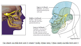
Trigeminal neuralgia
The trigeminal nerve transmits sensation from parts of the face to the brain and controls some of the muscles that are involved in chewing. Damage to this nerve causes repeated bursts of sharp, stabbing pain, known as trigeminal neuralgia, in the lip, gum, or cheek on one side of the face. Attacks may last for a few seconds or several minutes and may become more frequent over time. In severe cases, the pain may be so intense that the affected person is unable to do anything during the attack. Afterwards, the pain usually disappears completely. An attack may occur spontaneously or be triggered by certain facial movements, such as chewing, or by touching a trigger spot on the face. Attacks rarely occur at night.
Trigeminal neuralgia affects about 1 in 25,000 people in the UK. The disorder is most common in men over the age of 50. The cause of trigeminal neuralgia is usually unknown. However, in people under 50, symptoms may be an early sign of multiple sclerosis. Rarely, the nerve is compressed by a tumour or by a dilated blood vessel.
What may be done?
There are no specific tests to diagnose trigeminal neuralgia. Your doctor will examine you to rule out any other causes of facial pain, such as toothache or sinusitis. He or she may also arrange for you to have MRI to look for the presence of a tumour.
Your doctor may prescribe painkillers, such as paracetamol or ibuprofen. However, if the pain persists, your doctor may prescribe anticonvulsant drugs, such as carbamazapine, or certain antidepressants, all of which have been shown to be effective in treating trigeminal neuralgia. Unlike painkillers, which are taken only when the pain is present, both anticonvulsants and antidepressants need to be taken every day to prevent attacks.
If a tumour is found, surgery may be necessary to remove it. Surgery may also be used to separate the trigeminal nerve from a blood vessel if the vessel is compressing the nerve. Rarely, people who have persistent, severe pain that does not respond to drugs are offered treatment to numb the affected side of the face. For example, pain can be alleviated by using a heated probe to destroy the nerve. Alternatively, the trigeminal nerve may be cut, or alcohol may be injected into the nerve to deaden it. Trigeminal neuralgia will not recur after treatment to numb the face, but you will need to take care when consuming hot food or drinks because of the lack of sensation in your face.
Attacks of neuralgia may stop spontaneously, become more frequent, or persist unchanged for months or years. However, symptoms usually improve significantly with treatment.
Read more…
Trigeminal neuralgia affects about 1 in 25,000 people in the UK. The disorder is most common in men over the age of 50. The cause of trigeminal neuralgia is usually unknown. However, in people under 50, symptoms may be an early sign of multiple sclerosis. Rarely, the nerve is compressed by a tumour or by a dilated blood vessel.

What may be done?
There are no specific tests to diagnose trigeminal neuralgia. Your doctor will examine you to rule out any other causes of facial pain, such as toothache or sinusitis. He or she may also arrange for you to have MRI to look for the presence of a tumour.
Your doctor may prescribe painkillers, such as paracetamol or ibuprofen. However, if the pain persists, your doctor may prescribe anticonvulsant drugs, such as carbamazapine, or certain antidepressants, all of which have been shown to be effective in treating trigeminal neuralgia. Unlike painkillers, which are taken only when the pain is present, both anticonvulsants and antidepressants need to be taken every day to prevent attacks.
If a tumour is found, surgery may be necessary to remove it. Surgery may also be used to separate the trigeminal nerve from a blood vessel if the vessel is compressing the nerve. Rarely, people who have persistent, severe pain that does not respond to drugs are offered treatment to numb the affected side of the face. For example, pain can be alleviated by using a heated probe to destroy the nerve. Alternatively, the trigeminal nerve may be cut, or alcohol may be injected into the nerve to deaden it. Trigeminal neuralgia will not recur after treatment to numb the face, but you will need to take care when consuming hot food or drinks because of the lack of sensation in your face.
Attacks of neuralgia may stop spontaneously, become more frequent, or persist unchanged for months or years. However, symptoms usually improve significantly with treatment.
Nutritional neuropathies
In nutritional neuropathies, the peripheral nerves, which branch from the brain and spinal cord, are damaged by deficiencies of essential nutrients, particularly those of the vitamin B complex.
Worldwide, nutritional neuropathies are generally caused by malnutrition. In developed countries, such neuropathies are more commonly associated with excessive alcohol consumption. People who drink heavily often also have a poor diet, which can cause a vitamin B deficiency. In addition, alcohol may directly damage the peripheral nerves. A person who has been drinking heavily for 10 years or more has a greatly increased risk of developing a nutritional neuropathy.
Nutritional deficiencies may occur in people with eating disorders, such as anorexia nervosa. People with long-term conditions that affect absorption of nutrients from the intestines may also develop nutritional deficiencies.
What are the symptoms?
Nutritional neuropathies usually first affect the tips of the fingers and the toes. The symptoms appear gradually over several months or years and slowly progress up the limbs to the trunk. Symptoms may include:
- Loss of sensation.
- Pins and needles.
- Pain in the feet and/or the hands.
Walking may be clumsy as a result of the loss of sensation in the feet and legs.
If the motor nerves (nerves that stimulate the muscles) are affected, muscle weakness and wasting may develop and further affect the ability to walk.
What might be done?
Your doctor will examine you by checking your reflexes and your ability to feel sensation such as a pin prick. He or she may arrange for blood tests to look for a vitamin deficiency or for evidence of liver damage caused by excessive alcohol consumption. Your doctor may also arrange for tests that measure the extent of the nerve damage.
Nutritional neuropathies are treated by replacing the missing nutrients. This is done either by giving a course of oral supplements or, in some cases, by injections. Your doctor may also prescribe anticonvulsant drugs or antidepressants to help to control the symptoms. You may also be prescribed painkillers to relieve any discomfort. Nerve damage is usually irreversible, but with treatment the progression of the disease can usually be halted.
Read more…
Worldwide, nutritional neuropathies are generally caused by malnutrition. In developed countries, such neuropathies are more commonly associated with excessive alcohol consumption. People who drink heavily often also have a poor diet, which can cause a vitamin B deficiency. In addition, alcohol may directly damage the peripheral nerves. A person who has been drinking heavily for 10 years or more has a greatly increased risk of developing a nutritional neuropathy.
 |
| B vitamin supplement tablets - Wikipedia. |
What are the symptoms?
Nutritional neuropathies usually first affect the tips of the fingers and the toes. The symptoms appear gradually over several months or years and slowly progress up the limbs to the trunk. Symptoms may include:
- Loss of sensation.
- Pins and needles.
- Pain in the feet and/or the hands.
Walking may be clumsy as a result of the loss of sensation in the feet and legs.
If the motor nerves (nerves that stimulate the muscles) are affected, muscle weakness and wasting may develop and further affect the ability to walk.
What might be done?
Your doctor will examine you by checking your reflexes and your ability to feel sensation such as a pin prick. He or she may arrange for blood tests to look for a vitamin deficiency or for evidence of liver damage caused by excessive alcohol consumption. Your doctor may also arrange for tests that measure the extent of the nerve damage.
Nutritional neuropathies are treated by replacing the missing nutrients. This is done either by giving a course of oral supplements or, in some cases, by injections. Your doctor may also prescribe anticonvulsant drugs or antidepressants to help to control the symptoms. You may also be prescribed painkillers to relieve any discomfort. Nerve damage is usually irreversible, but with treatment the progression of the disease can usually be halted.
Diabetic neuropathy
In diabetic neuropathy, one or more of the peripheral nerves that branch from the brain and spinal cord to the rest of the body are damaged as a result of diabetes mellitus. Diabetic neuropathy is the most common cause of peripheral neuropathy in developed countries. If diabetes is poorly controlled, it results in high levels of glucose in the blood that damage the peripheral nerves directly and the blood vessels that supply them. Good control of diabetes reduces this risk by up to half.
About 3 in 10 people with diabetes mellitus have damage to one or more peripheral nerves, but only 1 of these 3 people develops significant symptoms.
People with diabetes mellitus who smoke increase the risk of damaging the blood vessels that supply the nerves,
What are the symptoms?
The symptoms of diabetic neuropathy usually develop slowly over a number of years. Rarely, they develop rapidly over days or weeks. Symptoms vary depending on which nerves are involved, but the feet are frequently affected. Less commonly, diabetic neuropathy may affect the larger nerves, mainly in the thighs. Symptoms may include:
- Pins and needles.
- Numbness.
- Pain, which is often worse at night.
- Muscle weakness and wasting.

In sensation is lost, a minor injury to the foot, such as rubbing by badly fitting shoes, may not be noticed. Slow healing due to poor blood supply may lead to infection. If left untreated, ulcers may develop and, in severe cases, gangrene occurs.
Eventually, diabetic neuropathy may also affect the autonomic nerves that regulate automatic body functions such as blood pressure control and digestion. Damage to these nerves causes symptoms such as dizziness when standing, diarrhea, and impotence.
What might be done?
Careful control of diabetes reduces the risk of developing diabetic neuropathy. However, if you develop symptoms of nerve damage, you should consult your doctor. He or she will probably be able to diagnose the condition from your symptoms. However, nerve conduction tests may need to be carried out in hospital to confirm which nerves are affected and to assess the severity of the damage. The goal of treatment of diabetic neuropathy is to prevent further nerve damage and the development of complications. Your doctor will help you to monitor your blood sugar level carefully and advise you about good foot care. For example, you should check your feet regular for cuts or abrasions, particularly if you have been wearing new shoes. You should avoid wearing open-toed sandals or walking barefoot. If you smoke, you should try to give up.
To relive pain, particularly at night, certain anticonvulsant drugs, such as carbamazepine and phenytoin, or an antidepressant drug, such as amitriptyline, may be prescribed.
What is the prognosis?
Good control of blood glucose levels in diabetes mellitus not only reduces the risk of developing diabetic neuropathy but may also half further progression of the disease. However, in most cases, nerve damage is irreversible.
Read more…
About 3 in 10 people with diabetes mellitus have damage to one or more peripheral nerves, but only 1 of these 3 people develops significant symptoms.
People with diabetes mellitus who smoke increase the risk of damaging the blood vessels that supply the nerves,
What are the symptoms?
The symptoms of diabetic neuropathy usually develop slowly over a number of years. Rarely, they develop rapidly over days or weeks. Symptoms vary depending on which nerves are involved, but the feet are frequently affected. Less commonly, diabetic neuropathy may affect the larger nerves, mainly in the thighs. Symptoms may include:
- Pins and needles.
- Numbness.
- Pain, which is often worse at night.
- Muscle weakness and wasting.
(tieuduong360.com)
In sensation is lost, a minor injury to the foot, such as rubbing by badly fitting shoes, may not be noticed. Slow healing due to poor blood supply may lead to infection. If left untreated, ulcers may develop and, in severe cases, gangrene occurs.
Eventually, diabetic neuropathy may also affect the autonomic nerves that regulate automatic body functions such as blood pressure control and digestion. Damage to these nerves causes symptoms such as dizziness when standing, diarrhea, and impotence.
What might be done?
Careful control of diabetes reduces the risk of developing diabetic neuropathy. However, if you develop symptoms of nerve damage, you should consult your doctor. He or she will probably be able to diagnose the condition from your symptoms. However, nerve conduction tests may need to be carried out in hospital to confirm which nerves are affected and to assess the severity of the damage. The goal of treatment of diabetic neuropathy is to prevent further nerve damage and the development of complications. Your doctor will help you to monitor your blood sugar level carefully and advise you about good foot care. For example, you should check your feet regular for cuts or abrasions, particularly if you have been wearing new shoes. You should avoid wearing open-toed sandals or walking barefoot. If you smoke, you should try to give up.
To relive pain, particularly at night, certain anticonvulsant drugs, such as carbamazepine and phenytoin, or an antidepressant drug, such as amitriptyline, may be prescribed.
What is the prognosis?
Good control of blood glucose levels in diabetes mellitus not only reduces the risk of developing diabetic neuropathy but may also half further progression of the disease. However, in most cases, nerve damage is irreversible.
Peripheral neuropathies
Disorders of the peripheral nerves, the nerves that branch from the brain and spinal cord to the rest of the body, are called neuropathies. Depending on the nerves affected, peripheral neuropathies may affect sensation, movement, or automatic functions, such as bladder control. Rarely, a peripheral neuropathy may be life-threatening.
What are the causes?
In developed countries, the most common cause of damage to the peripheral nerves is diabetes mellitus. Vitamin B complex deficiencies and some nutritional disorders may also result in nerve damage. In the developed world, nutritional neuropathy is often the result of a poor diet in people who abuse alcohol. Drinking too much alcohol may also damage peripheral nerves directly.
Damage to a single nerve may occur as a result of an injury or because of compression. For example, in carpal tunnel syndrome, the median nerve, which supplies that part of the hand, is compressed at the wrist.
Neuropathy may also be associated with an infection, such as Hansen’s disease or HIV infection. Guilain-Barre syndrome, a neuropathy that is rapidly progressive, is caused by an abnormal immune response that sometimes occurs after an infection.
Autoimmune disorders such as systemic lupus erythematosus, in which the immune system attacks the body’s tissues, may cause nerve damage occasionally, a disorder such as polyarteritis nodosamay damage nerves by causing inflammation of the blood vessels that supply them. Neuropathy may also result from certain cancers, particularly primary lung cancer and lymphoma. Occasionally, neuropathy is caused by amyloidosis, in which an abnormal protein is deposited in the body.
Some drugs, such as isoniazid, may cause nerve damage, as may exposure to certain toxic substances, such as lead. In some cases, the cause is unknown.
What are the types?
Peripheral neuropathies may affect the nerves that transmit sensory information (sensory nerves), the nerves that stimulate the muscles (motornerves), and/or the nerves that control automatic functions (autonomic nerves).
Sensory nerve neuropathies: these neuropathies first affect the hands and feet and the spread towards the centre of the body. The symptoms may include tingling, pain, and numbness in the affected area. If the fingertips are numb, everyday tasks may become difficult. This type of neuropathy is most often caused by nutritional disorders or drugs.
Motor nerve neuropathies: if the motor nerves are damaged, the muscles they supply become weak, and wasting occurs eventually. In severe cases, mobility may become restricted, and very rarely, breathing may have to be assisted by mechanical ventilation. Lead poisoning may result in a neuropathy that affects the motor nerves only.
Autonomic nerve neuropathies: a neuropathy that is affecting one or more autonomic nerves may result in constipation, fainting due to low blood pressure, diarrhoea, urinary in continence, or impotence. This type of neuropathy is often caused by long-standing diabetes mellitus.
What might be done?
Your doctor may be able to tell which nerves are affected from your symptoms and an examination. If the cause of your neuropathy is not clear, he or she will probably arrange for blood tests to look for evidence of an underlying disorder, such as nutritional deficiencies or an autoimmune disorder. If Mere is evidence of compression of a nerve, you may also have CT scanning or MRI to assess the severity and extent of nerve damage. Special tests to assess the function of the nerves may also be carried out.
The treatment of a peripheral neuropathy depends on the cause and the type of nerve affected. For example, careful control of diabetes mellitus may keep diabetic neuropathy from worsening, and vitamin B complex injections may help a nutritional neuropathy. If motor nerves are affected, you may have physiotherapy to help to maintain muscle tone. Wearing a foot splint may assist walking. Sometimes, the underlying cause can be treated, but long-standing nerve damage may be irreversible.
TEST NERVE AND MUSLCE ELECTRICAL TESTS
Nerve and muscle electrical test consist of nerve conduction studies and electromyography (EMG). Nerve conduction studies are used to assess how well a nerve is conducting electrical impulses. They are often followed by EMG to see whether symptoms, such as weakness, are due to a disorder of the muscle or the nerve supplying it. Both tests are usually done on an outpatient basis. Each takes about 15 minutes and may cause discomfort.
Nerve conduction studies
Nerve conduction studies are carried out to assess nerve damage in disorders such as peripheral neuropathies. A nerve is stimulated by an electrical impulse, and the response to the stimulus and the speed at which this response travels along the nerve indicates whether the nerve is damaged and the nature and extent of the damage.
Electromyography
EMG is used to differentiate between nerve and muscle disorders and to diagnose disorders such as muscular dystrophy. A fine needle is used to record the electrical activity of a muscle at rest and when contracting. The results are recorded on a trace.
Read more…
What are the causes?
In developed countries, the most common cause of damage to the peripheral nerves is diabetes mellitus. Vitamin B complex deficiencies and some nutritional disorders may also result in nerve damage. In the developed world, nutritional neuropathy is often the result of a poor diet in people who abuse alcohol. Drinking too much alcohol may also damage peripheral nerves directly.
Damage to a single nerve may occur as a result of an injury or because of compression. For example, in carpal tunnel syndrome, the median nerve, which supplies that part of the hand, is compressed at the wrist.
Neuropathy may also be associated with an infection, such as Hansen’s disease or HIV infection. Guilain-Barre syndrome, a neuropathy that is rapidly progressive, is caused by an abnormal immune response that sometimes occurs after an infection.
Autoimmune disorders such as systemic lupus erythematosus, in which the immune system attacks the body’s tissues, may cause nerve damage occasionally, a disorder such as polyarteritis nodosamay damage nerves by causing inflammation of the blood vessels that supply them. Neuropathy may also result from certain cancers, particularly primary lung cancer and lymphoma. Occasionally, neuropathy is caused by amyloidosis, in which an abnormal protein is deposited in the body.
Some drugs, such as isoniazid, may cause nerve damage, as may exposure to certain toxic substances, such as lead. In some cases, the cause is unknown.
 |
A painless ulcer developing under the big toe joint due to peripheral neuropathy |
What are the types?
Peripheral neuropathies may affect the nerves that transmit sensory information (sensory nerves), the nerves that stimulate the muscles (motornerves), and/or the nerves that control automatic functions (autonomic nerves).
Sensory nerve neuropathies: these neuropathies first affect the hands and feet and the spread towards the centre of the body. The symptoms may include tingling, pain, and numbness in the affected area. If the fingertips are numb, everyday tasks may become difficult. This type of neuropathy is most often caused by nutritional disorders or drugs.
Motor nerve neuropathies: if the motor nerves are damaged, the muscles they supply become weak, and wasting occurs eventually. In severe cases, mobility may become restricted, and very rarely, breathing may have to be assisted by mechanical ventilation. Lead poisoning may result in a neuropathy that affects the motor nerves only.
Autonomic nerve neuropathies: a neuropathy that is affecting one or more autonomic nerves may result in constipation, fainting due to low blood pressure, diarrhoea, urinary in continence, or impotence. This type of neuropathy is often caused by long-standing diabetes mellitus.
What might be done?
Your doctor may be able to tell which nerves are affected from your symptoms and an examination. If the cause of your neuropathy is not clear, he or she will probably arrange for blood tests to look for evidence of an underlying disorder, such as nutritional deficiencies or an autoimmune disorder. If Mere is evidence of compression of a nerve, you may also have CT scanning or MRI to assess the severity and extent of nerve damage. Special tests to assess the function of the nerves may also be carried out.
The treatment of a peripheral neuropathy depends on the cause and the type of nerve affected. For example, careful control of diabetes mellitus may keep diabetic neuropathy from worsening, and vitamin B complex injections may help a nutritional neuropathy. If motor nerves are affected, you may have physiotherapy to help to maintain muscle tone. Wearing a foot splint may assist walking. Sometimes, the underlying cause can be treated, but long-standing nerve damage may be irreversible.
TEST NERVE AND MUSLCE ELECTRICAL TESTS
Nerve and muscle electrical test consist of nerve conduction studies and electromyography (EMG). Nerve conduction studies are used to assess how well a nerve is conducting electrical impulses. They are often followed by EMG to see whether symptoms, such as weakness, are due to a disorder of the muscle or the nerve supplying it. Both tests are usually done on an outpatient basis. Each takes about 15 minutes and may cause discomfort.
Nerve conduction studies
Nerve conduction studies are carried out to assess nerve damage in disorders such as peripheral neuropathies. A nerve is stimulated by an electrical impulse, and the response to the stimulus and the speed at which this response travels along the nerve indicates whether the nerve is damaged and the nature and extent of the damage.
Electromyography
EMG is used to differentiate between nerve and muscle disorders and to diagnose disorders such as muscular dystrophy. A fine needle is used to record the electrical activity of a muscle at rest and when contracting. The results are recorded on a trace.
Multiple sclerosis
Multiple sclerosis (MS) is the most common nervous system disorder affecting young adults. In this condition, nerves in the brain and spinal cord are progressively damaged, causing a wide range of symptoms that affect sensation, movement, body functions, and balance. Specific symptoms may relate to the particular areas that are damaged and vary in severity between individuals. For example, damage to the optic nerve may cause blurred vision. If nerve fibres in the spinal cord are affected, it may cause weakness and heaviness in the legs or arms. Damage to nerves in the brain stem, the area of the brain that connects to the spinal cord, may affect balance.
In many people with MS, symptoms occur intermittently and there may be long periods of remission. However, some people have chronic (long-term) symptoms that gradually get worse.
In the UK, about 85,000 people are affected by MS. People who have a close relative with MS are more likely to develop the disorder. The condition is much more common in the northern hemisphere, which suggests that environmental factors also play a part. MS is more common in females and the disorder is more likely develop between early adulthood and middle age.
What are the causes?
MS is an autoimmune disorder, in which the body’s immune system attacks its own tissue, in this case those of the nervous system. Many nerves in the brain and spinal cord are covered by a protective insulating sheath of material called myelin. In MS, small areas of myelin are damaged, leaving holes in the sheath, a process known as demyelination. Once the myelin sheath has been damaged, impulses cannot be conducted normally along nerves to and from the brain and spinal cord. At first, the damage may be limited to only one nerve, but myelin covering other nerves may become damaged over time. Eventually, damaged parches of myelin insulation are replaced by scar tissue.
It is thought that MS may be triggered by external factors such as a viral infection during childhood in genetically susceptible individuals.
What are the types?
There are two types of MS. In the most common, known as relapsing remitting MS, symptoms last for days or weeks and then clear up for months or even years. However, some symptoms may eventually persist between the attacks. About 3 in 10 people with MS have a type known as chronic-progressive MS, in which there is a gradual worsening of symptoms with no remission.
A person with relapsing-remitting MS may go on to develop chronic-progressive MS.
What are the symptoms?

Symptoms may occur singly in the initial stages and in combination as the disorder progresses. They may include:
- Blurred vision.
- Numbness or tingling in any part of the body.
- Tiredness, which may be persistent.
- Weakness and a feeling of heaviness in the legs or arms.
- Problems with coordination and balance, such as an unsteady gait.
Stress and heat sometimes make symptoms worse. About half of the people who have MS find it hard to concentrate and experience memory lapses. Depression is common. Later in the course of the disease, some people with muscle weakness develop painful muscle spasms. Spinal cord damage can lead to urinary incontinence, and men may have increasing difficulty in achieving an erection. Eventually, damage to myelin covering nerves in the spinal cord may cause partial paralysis, and an affected person may need a wheelchair.
How is it diagnosed?
There is no single test to diagnose MS, and, because symptoms are so wide-ranging, a diagnosis is only made once other possible causes of the symptoms have been excluded. Your doctor will take your medical history and carry out a physical examination. If you are having visual problems, such as blurred vision, you may be referred to an ophthalmologist, who will assess the optic nerve, which is commonly affected in the early stages of the disorder. Your doctor may arrange for tests to find out how quickly your brain receives messages when particular nerves are stimulated. The most common test measures damage to the visual pathways. You will probably also have an imaging test of the brain, such as MRI, to see if there are areas of demyelination.
Your doctor may arrange for a lumbar puncture, a procedure in which a small amount of the fluid that surrounds the spinal cord is removed for microscopic analysis. Abnormalities in this fluid may confirm the diagnosis.
What is the treatment?
There is no cure for MS, but if you have relapsing-remitting MS, interferon beta may help to lengthen remission periods and shorten the length of attacks. Your doctor may also prescribe corticosteroids to shorten the duration of a relapse. However, at present, there is no specific treatment to halt the progression of chronic-progressive MS.
Many of the more common symptoms that occur in all types of MS can be relieved by drugs. For example, your doctor may treat muscle spasms with a drug that relaxes muscles. Similarly, incontinence can often be improved by drugs. Problems in getting an erection may be helped by a drugs treatment such as sildenafil. If you have mobility problems, your doctor may arrange for you to have physiotherapy. Occupational therapy may make day-to-day activities easier.
What can I do?
If you are diagnosed with MS, you and your family will need time and possibly counseling to come to terms with the disorder. You should minimize stress in your life and avoid exposure to high temperatures if hear tends to make your symptoms worse. Regular, gentle exercise, such as swimming, will help to keep your muscle strong without the risk of overstraining them.
The progression of MS is extremely variable, but people who are older when the disease first develops tend to fare less well. About 7 in 10 people with MS have active lives with long periods of remission between relapses. However, some people, particularly those with chronic-progressive MS, become increasingly disabled. Half of all people with MS are still leading active lives 10 years after diagnosis, and the average lifespan from diagnosis is 25-30 years.
VISUAL EVOKED RESPONSES
A visual evoked response test measure the function of the optic nerve, the nerve that transmits messages from the eye to the brain. The test is most often used to in the diagnosis of multiple sclerosis and can detect abnormalities even if visual symptoms are not apparent. The test records brain activity in response to a visual stimulus to find out the speed at which messages from the eye reach the brain. The test takes 20-30 minutes.
Read more…
In many people with MS, symptoms occur intermittently and there may be long periods of remission. However, some people have chronic (long-term) symptoms that gradually get worse.
In the UK, about 85,000 people are affected by MS. People who have a close relative with MS are more likely to develop the disorder. The condition is much more common in the northern hemisphere, which suggests that environmental factors also play a part. MS is more common in females and the disorder is more likely develop between early adulthood and middle age.
What are the causes?
MS is an autoimmune disorder, in which the body’s immune system attacks its own tissue, in this case those of the nervous system. Many nerves in the brain and spinal cord are covered by a protective insulating sheath of material called myelin. In MS, small areas of myelin are damaged, leaving holes in the sheath, a process known as demyelination. Once the myelin sheath has been damaged, impulses cannot be conducted normally along nerves to and from the brain and spinal cord. At first, the damage may be limited to only one nerve, but myelin covering other nerves may become damaged over time. Eventually, damaged parches of myelin insulation are replaced by scar tissue.
It is thought that MS may be triggered by external factors such as a viral infection during childhood in genetically susceptible individuals.
What are the types?
There are two types of MS. In the most common, known as relapsing remitting MS, symptoms last for days or weeks and then clear up for months or even years. However, some symptoms may eventually persist between the attacks. About 3 in 10 people with MS have a type known as chronic-progressive MS, in which there is a gradual worsening of symptoms with no remission.
A person with relapsing-remitting MS may go on to develop chronic-progressive MS.
What are the symptoms?
Symptoms may occur singly in the initial stages and in combination as the disorder progresses. They may include:
- Blurred vision.
- Numbness or tingling in any part of the body.
- Tiredness, which may be persistent.
- Weakness and a feeling of heaviness in the legs or arms.
- Problems with coordination and balance, such as an unsteady gait.
Stress and heat sometimes make symptoms worse. About half of the people who have MS find it hard to concentrate and experience memory lapses. Depression is common. Later in the course of the disease, some people with muscle weakness develop painful muscle spasms. Spinal cord damage can lead to urinary incontinence, and men may have increasing difficulty in achieving an erection. Eventually, damage to myelin covering nerves in the spinal cord may cause partial paralysis, and an affected person may need a wheelchair.
How is it diagnosed?
There is no single test to diagnose MS, and, because symptoms are so wide-ranging, a diagnosis is only made once other possible causes of the symptoms have been excluded. Your doctor will take your medical history and carry out a physical examination. If you are having visual problems, such as blurred vision, you may be referred to an ophthalmologist, who will assess the optic nerve, which is commonly affected in the early stages of the disorder. Your doctor may arrange for tests to find out how quickly your brain receives messages when particular nerves are stimulated. The most common test measures damage to the visual pathways. You will probably also have an imaging test of the brain, such as MRI, to see if there are areas of demyelination.
Your doctor may arrange for a lumbar puncture, a procedure in which a small amount of the fluid that surrounds the spinal cord is removed for microscopic analysis. Abnormalities in this fluid may confirm the diagnosis.
What is the treatment?
There is no cure for MS, but if you have relapsing-remitting MS, interferon beta may help to lengthen remission periods and shorten the length of attacks. Your doctor may also prescribe corticosteroids to shorten the duration of a relapse. However, at present, there is no specific treatment to halt the progression of chronic-progressive MS.
Many of the more common symptoms that occur in all types of MS can be relieved by drugs. For example, your doctor may treat muscle spasms with a drug that relaxes muscles. Similarly, incontinence can often be improved by drugs. Problems in getting an erection may be helped by a drugs treatment such as sildenafil. If you have mobility problems, your doctor may arrange for you to have physiotherapy. Occupational therapy may make day-to-day activities easier.
What can I do?
If you are diagnosed with MS, you and your family will need time and possibly counseling to come to terms with the disorder. You should minimize stress in your life and avoid exposure to high temperatures if hear tends to make your symptoms worse. Regular, gentle exercise, such as swimming, will help to keep your muscle strong without the risk of overstraining them.
The progression of MS is extremely variable, but people who are older when the disease first develops tend to fare less well. About 7 in 10 people with MS have active lives with long periods of remission between relapses. However, some people, particularly those with chronic-progressive MS, become increasingly disabled. Half of all people with MS are still leading active lives 10 years after diagnosis, and the average lifespan from diagnosis is 25-30 years.
VISUAL EVOKED RESPONSES
A visual evoked response test measure the function of the optic nerve, the nerve that transmits messages from the eye to the brain. The test is most often used to in the diagnosis of multiple sclerosis and can detect abnormalities even if visual symptoms are not apparent. The test records brain activity in response to a visual stimulus to find out the speed at which messages from the eye reach the brain. The test takes 20-30 minutes.
Motor neuron disease
Motor neuron disease is rare, affecting 1-2 people in every 100,000 worldwide each year. In this disease, also known as amyotrophic lateral sclerosis, degeneration of the nerves involved in muscular activity results in progressive wasting of the muscles and weakness. There are several types of motor neuron disease. Some affect mainly the spinal nerves, while other types also affect the brain. The condition is not painful, does not affect bowel or bladder function, and does not usually affect the intellect or the senses, such as sight.
The cause of motor neuron disease is unknown. Genetic factors are involved because, in about 1 in 10 cases, the disease runs in the family. The disease is slightly more common in men and usually develops after the age of 40.
What are the symptoms?
Initially, weakness and wasting develop over a few months and usually affect the muscles of the hands, arms, or legs. Other early symptoms may include:
- Twitching movements in the muscles.
- Stiffness and muscle cramps.
- Difficulty in carrying out twisting movements, such as unscrewing bottle tops and turning keys.
As the disease progresses, other symptoms may include:
- Dragging one foot or a tendency to stumble when walking.
- Difficulty in climbing stairs or getting up from low chairs.
Less commonly, the muscles of the mouth and throat are involved, and may cause slurred speech, hoarseness, and difficulty in swallowing.
An affected person may have mood swings and may become anxious and depressed. If the muscles involved in breathing and swallowing are affected, small particles of food may enter the lungs and cause recurrent chest infections and possibly pneumonia. The head may fall forwards because the muscles in the neck are too weak to support it. Eventually, weakness of the muscles that control respiration may cause difficulty in breathing.
How is diagnosed?
There is no specific test to diagnose motor neuron disease. However, electromyography may be carried out to detect a decrease in electrical activity in the muscles. Additional tests may be used to exclude other possible causes of the symptoms. For example, MRI or CT scanning of the brain and neck may be used to exclude a local problem such as a tumour or cervical spondylosis.
What is the treatment?
At present, no treatment can significantly slow down the progression of motor neuron disease, although a new drug called riluzole may have a small effect. Treatment for symptoms may include antidepressants to relieve depression and antibiotics to treat chest infections. If the person is having difficulty in swallowing, a gastrostomy maybe created surgically. This is an opening through which a permanent feeding tube is inserted directly into the stomach or the small intestine.
Usually, a team of specialists provide support and care for an affected person and members of the family. Counseling may be offered to both. The person affected by the disease may have physiotherapy to keep joints and muscles supple and may be given aids to help with activities such as eating and walking. A speech therapist can supply communication aids to help with speech difficulties and advise on swallowing problems. Joining a self-help group is often helpful to person with motor neuron disease and his or her family.
The outlook for motor neuron disease is variable, with approximately 2 in 10 affected people alive 5 years after diagnosis. About 1 in 10 affected people survives more than 10 years.
Read more…
 |
Steven Hawkins is the one of few people to
ever live passed 5 years with MND.
He has lived for around 40 years |
The cause of motor neuron disease is unknown. Genetic factors are involved because, in about 1 in 10 cases, the disease runs in the family. The disease is slightly more common in men and usually develops after the age of 40.
What are the symptoms?
Initially, weakness and wasting develop over a few months and usually affect the muscles of the hands, arms, or legs. Other early symptoms may include:
- Twitching movements in the muscles.
- Stiffness and muscle cramps.
- Difficulty in carrying out twisting movements, such as unscrewing bottle tops and turning keys.
As the disease progresses, other symptoms may include:
- Dragging one foot or a tendency to stumble when walking.
- Difficulty in climbing stairs or getting up from low chairs.
Less commonly, the muscles of the mouth and throat are involved, and may cause slurred speech, hoarseness, and difficulty in swallowing.
An affected person may have mood swings and may become anxious and depressed. If the muscles involved in breathing and swallowing are affected, small particles of food may enter the lungs and cause recurrent chest infections and possibly pneumonia. The head may fall forwards because the muscles in the neck are too weak to support it. Eventually, weakness of the muscles that control respiration may cause difficulty in breathing.
How is diagnosed?
There is no specific test to diagnose motor neuron disease. However, electromyography may be carried out to detect a decrease in electrical activity in the muscles. Additional tests may be used to exclude other possible causes of the symptoms. For example, MRI or CT scanning of the brain and neck may be used to exclude a local problem such as a tumour or cervical spondylosis.
What is the treatment?
At present, no treatment can significantly slow down the progression of motor neuron disease, although a new drug called riluzole may have a small effect. Treatment for symptoms may include antidepressants to relieve depression and antibiotics to treat chest infections. If the person is having difficulty in swallowing, a gastrostomy maybe created surgically. This is an opening through which a permanent feeding tube is inserted directly into the stomach or the small intestine.
Usually, a team of specialists provide support and care for an affected person and members of the family. Counseling may be offered to both. The person affected by the disease may have physiotherapy to keep joints and muscles supple and may be given aids to help with activities such as eating and walking. A speech therapist can supply communication aids to help with speech difficulties and advise on swallowing problems. Joining a self-help group is often helpful to person with motor neuron disease and his or her family.
The outlook for motor neuron disease is variable, with approximately 2 in 10 affected people alive 5 years after diagnosis. About 1 in 10 affected people survives more than 10 years.
Creutzfeldt-Jakob disease
Creutzfeldt-Jakob disease (CJD) is an extremely rare condition in which brain tissue is progressively destroyed by an unusual infectious agent. The disorder leads to a general decline in all areas of mental, and physical ability and ultimately to death. CJD affects about one person in a million each year worldwide.
What is the cause?
CJD is caused by an infectious agent known as a prion, which replicates in the brain and causes brain damage. One type of CJD, accounting for 15 in 100 cases, has been found to run in families.
Most people who develop CJD are over the age of 50. Usually, the source of the infection is unknown, but in about 1 in 20 people it can be traced to earlier treatment with products derived from human tissue. Before the use of artificial growth hormones to treat growth disorders, human growth hormone injections were one source of infection.

In the mid 1990s, a new, rare variant of CJD that affects people in their teens or 20s was discovered in the UK. By the year 2000, about 50 cases had been reported. This variant is believed to be linked with eating contaminated meat from cattle with a disease called bovine spongiform encephalopathy (BSE).
What are the symptoms?
It is thought that is present for 2-15 years before symptoms begin to develop gradually. They may include:
- Depression.
- Poor memory.
- Unsteadiness and poor coordination.
Other symptoms develop as the condition progresses and include:
- Sudden muscle contractions.
- Seizures.
- Weakness or paralysis on one side of the body.
- Progressive dementia.
- Impaired vision.
In the later stages CJD, a person may be unable to move and talk. Those who are confined to bed are prone to serious lung infections.
What might be done?
CJD is usually diagnosed from a person’s symptoms because no specific test is yet available. Anyone suspected of having CJD will have extensive tests, such as MRI, to exclude other treatable causes, and EEG to look for characteristic changes in electrical activity in the brain. A brain biopsy, in which a small piece of tissue is surgically removed for examination, may be performed. There is no cure for CJD, but drugs can relieve some symptoms. For example, symptoms of depression may be treated with antidepressant drugs, and muscle contractions may be controlled by muscle relaxant drugs. However, the disorder is usually fatal within 3 years.
Read more…
What is the cause?
CJD is caused by an infectious agent known as a prion, which replicates in the brain and causes brain damage. One type of CJD, accounting for 15 in 100 cases, has been found to run in families.
Most people who develop CJD are over the age of 50. Usually, the source of the infection is unknown, but in about 1 in 20 people it can be traced to earlier treatment with products derived from human tissue. Before the use of artificial growth hormones to treat growth disorders, human growth hormone injections were one source of infection.
In the mid 1990s, a new, rare variant of CJD that affects people in their teens or 20s was discovered in the UK. By the year 2000, about 50 cases had been reported. This variant is believed to be linked with eating contaminated meat from cattle with a disease called bovine spongiform encephalopathy (BSE).
What are the symptoms?
It is thought that is present for 2-15 years before symptoms begin to develop gradually. They may include:
- Depression.
- Poor memory.
- Unsteadiness and poor coordination.
Other symptoms develop as the condition progresses and include:
- Sudden muscle contractions.
- Seizures.
- Weakness or paralysis on one side of the body.
- Progressive dementia.
- Impaired vision.
In the later stages CJD, a person may be unable to move and talk. Those who are confined to bed are prone to serious lung infections.
What might be done?
CJD is usually diagnosed from a person’s symptoms because no specific test is yet available. Anyone suspected of having CJD will have extensive tests, such as MRI, to exclude other treatable causes, and EEG to look for characteristic changes in electrical activity in the brain. A brain biopsy, in which a small piece of tissue is surgically removed for examination, may be performed. There is no cure for CJD, but drugs can relieve some symptoms. For example, symptoms of depression may be treated with antidepressant drugs, and muscle contractions may be controlled by muscle relaxant drugs. However, the disorder is usually fatal within 3 years.
Parkinson’s disease and Parkinsonism
Parkinson’s disease results from degeneration of cells in a part of the brain called the basal ganglia, which controls the smoothness of muscle movements. Normally, these cells produce a neurotransmitter (a chemical that transmits nerve impulses) called dopamine, which acts with acetylcholine, an other neuro-transmitter, to fine-tune muscle control. In Parkinson’s disease, the level of dopamine relative to acetylcholine is reduced, adversely affecting muscle control.
About 1 in 200 people in the UJ has Parkinson’s disease. The disorder tends to occur after the age of 60 and is more common in men.
Parkinsonism is the term used for symptoms of Parkinson’s disease when they are due to an underlying disorder or certain drugs. Repeated head injuries may cause Parkinsonism, as may some antipsychotic drugs used to treat severe psychiatric illness.

What are the symptoms?
The main symptoms of Parkinson’s disease develop gradually over months or even years. Parkinsonism may have a gradual or sudden onset depending on the cause. Symptoms include:
- Tremor of on hand, arm or leg, usually when resting, that later occurs on both sides.
- Muscle stiffness, making it difficult to start moving.
- Slowness of movement.
- Shuffling walk with loss of arm swing.
- Expressionless or mask-like face.
- Stooped posture.
As the disease progresses, stiffness, immobility, and constant trembling of the hands may make some daily tasks difficult to perform. Speech may become slow and hesitant, and swallowing may be difficult. Many people with the disorder develop depression. About 3 in 10 people with the disease eventually develop dementia.
How is it diagnosed?
Since Parkinson’s disease begins gradually, it is often not possible to diagnose the condition immediately. Your doctor will examine you and may arrange tests such as CT scanning or MRI to exclude other possible causes. If a specific underlying disorder is found to be causing your symptoms, you will be diagnosed as having Parkinsonism rather than Parkinson’s disease.
How might the doctor treat it?
There is no specific cure for Parkinson’s disease, but drugs physical treatments, and more rarely, surgery can relieve symptoms. If you have Parkinsonism due to medications, your doctor may change your drugs. Symptoms the usually disappear within 8 weeks. If the symptoms persist, you may need to be treated with anti-parkinsonism drugs.
Drug treatment: in the early stages of Parkinson’s disease when symptoms are mild, treatment may not be necessary because drugs can not change the progression of the disease. Later on, drugs are used to relieve symptoms and reduce disability by correcting chemical imbalances in the brain, either by boosting dopamine levels or by blocking some the effects of acetylcholine or a combination of both. The choice and dosage of drugs is tailored to individual’s particular problems and adjusted to reduce side effects.
Often, an anti-cholinergic drug such as trihexyphenidyl (benzhexol) is given initially to reduce shaking and stiffness. Anti-cholinergic drugs can be effective for several years, but they may cause dry mouth, burred vision and difficulty in passing urine.
The main treatment for Parkinson’s disease is with levodopa, which boosts the level of dopamine within the brain. Levodopa controls major symptoms such as mobility problems but may initially cause side effects, such as nausea and vomiting. For this reason, the drug is increased gradually and is usually prescribed with other drugs, such as carbidopa or benserazide, which reduce the amount of levodopa that is needed. However, some people develop involuntary jerky or writhing movements as a side effect of this treatment. Levodopa is usually effective for 2-5 years, but long-term use may be associated with abrupt changes in symptoms known as the “one-off” effect of levodopa. The affected person has periods of normal mobility punctuated by unpredictable episodes of weakness and difficulty in moving.
When levodopa is no longer effective, seligiline, a drugs that slightly increases dopamine activity in the brain, may be tried, and later, other drugs, including dopamine agonists such s bromocriptine, may be used. Such drugs may cause confusion, hallucinations, and occasionally, aggressive behaviour. Although there is no wholly successful therapy for symptoms of this disease, new drugs are always being developed.
Physical treatment: the doctor may arrange for physiotherapy to help with mobility problems or speech therapy for speech and swallowing problems. If you are finding it difficult to cope at home, an occupational therapist may suggest changes, such as installing hand-rails to make it easier for you to move around.
Surgical treatment: younger people who are otherwise in good health may have surgery if the tremor cannot be controlled by drugs. Surgery involves destroying a part of the brain tissue responsible for the tremor. Therapies still being assessed include replacement of damaged brain cells with transplanted tissue and deep brain stimulation with electrical impulses to reduce tremor. Deep brain stimulation, in particular, holds promise as a future treatment.
What can I do?
It is important to pay attention to your general health. Taking a walk each day and doing simple stretching exercises will help you to maintain strength and mobility. You should also rest during they day to avoid getting tired. Emotional and practical help from family, friends, and support groups is important.
What is the prognosis?
The course of the Parkinson’s disease is variable, but drugs can be effective in treating the symptoms and improving the quality of life. People can lead active lives for many years after being diagnosed. However, most people with the disorder need daily help eventually, and their symptoms may be increasingly hard to control with drugs.
Read more…
About 1 in 200 people in the UJ has Parkinson’s disease. The disorder tends to occur after the age of 60 and is more common in men.
Parkinsonism is the term used for symptoms of Parkinson’s disease when they are due to an underlying disorder or certain drugs. Repeated head injuries may cause Parkinsonism, as may some antipsychotic drugs used to treat severe psychiatric illness.

What are the symptoms?
The main symptoms of Parkinson’s disease develop gradually over months or even years. Parkinsonism may have a gradual or sudden onset depending on the cause. Symptoms include:
- Tremor of on hand, arm or leg, usually when resting, that later occurs on both sides.
- Muscle stiffness, making it difficult to start moving.
- Slowness of movement.
- Shuffling walk with loss of arm swing.
- Expressionless or mask-like face.
- Stooped posture.
As the disease progresses, stiffness, immobility, and constant trembling of the hands may make some daily tasks difficult to perform. Speech may become slow and hesitant, and swallowing may be difficult. Many people with the disorder develop depression. About 3 in 10 people with the disease eventually develop dementia.
How is it diagnosed?
Since Parkinson’s disease begins gradually, it is often not possible to diagnose the condition immediately. Your doctor will examine you and may arrange tests such as CT scanning or MRI to exclude other possible causes. If a specific underlying disorder is found to be causing your symptoms, you will be diagnosed as having Parkinsonism rather than Parkinson’s disease.
How might the doctor treat it?
There is no specific cure for Parkinson’s disease, but drugs physical treatments, and more rarely, surgery can relieve symptoms. If you have Parkinsonism due to medications, your doctor may change your drugs. Symptoms the usually disappear within 8 weeks. If the symptoms persist, you may need to be treated with anti-parkinsonism drugs.
Drug treatment: in the early stages of Parkinson’s disease when symptoms are mild, treatment may not be necessary because drugs can not change the progression of the disease. Later on, drugs are used to relieve symptoms and reduce disability by correcting chemical imbalances in the brain, either by boosting dopamine levels or by blocking some the effects of acetylcholine or a combination of both. The choice and dosage of drugs is tailored to individual’s particular problems and adjusted to reduce side effects.
Often, an anti-cholinergic drug such as trihexyphenidyl (benzhexol) is given initially to reduce shaking and stiffness. Anti-cholinergic drugs can be effective for several years, but they may cause dry mouth, burred vision and difficulty in passing urine.
The main treatment for Parkinson’s disease is with levodopa, which boosts the level of dopamine within the brain. Levodopa controls major symptoms such as mobility problems but may initially cause side effects, such as nausea and vomiting. For this reason, the drug is increased gradually and is usually prescribed with other drugs, such as carbidopa or benserazide, which reduce the amount of levodopa that is needed. However, some people develop involuntary jerky or writhing movements as a side effect of this treatment. Levodopa is usually effective for 2-5 years, but long-term use may be associated with abrupt changes in symptoms known as the “one-off” effect of levodopa. The affected person has periods of normal mobility punctuated by unpredictable episodes of weakness and difficulty in moving.
When levodopa is no longer effective, seligiline, a drugs that slightly increases dopamine activity in the brain, may be tried, and later, other drugs, including dopamine agonists such s bromocriptine, may be used. Such drugs may cause confusion, hallucinations, and occasionally, aggressive behaviour. Although there is no wholly successful therapy for symptoms of this disease, new drugs are always being developed.
Physical treatment: the doctor may arrange for physiotherapy to help with mobility problems or speech therapy for speech and swallowing problems. If you are finding it difficult to cope at home, an occupational therapist may suggest changes, such as installing hand-rails to make it easier for you to move around.
Surgical treatment: younger people who are otherwise in good health may have surgery if the tremor cannot be controlled by drugs. Surgery involves destroying a part of the brain tissue responsible for the tremor. Therapies still being assessed include replacement of damaged brain cells with transplanted tissue and deep brain stimulation with electrical impulses to reduce tremor. Deep brain stimulation, in particular, holds promise as a future treatment.
What can I do?
It is important to pay attention to your general health. Taking a walk each day and doing simple stretching exercises will help you to maintain strength and mobility. You should also rest during they day to avoid getting tired. Emotional and practical help from family, friends, and support groups is important.
What is the prognosis?
The course of the Parkinson’s disease is variable, but drugs can be effective in treating the symptoms and improving the quality of life. People can lead active lives for many years after being diagnosed. However, most people with the disorder need daily help eventually, and their symptoms may be increasingly hard to control with drugs.
Huntington’s disease
Huntington’s disease is an inherited disorder that causes degeneration of a particular part of the brain. Also known as Huntington’s chorea, the condition causes jerky, involuntary movements, clumsiness, and progressive dementia. Huntington’s disease is rare, affecting only about 6,000 people in the UK. The symptoms commonly develop between the ages of 30 and 50.
Huntington’s disease is caused by an abnormal dominant gene. To develop the disease, an affected person has to inherit the abnormal gene from only one parent. People who have the gene have a 1 in 2 chance of passing it on to each of their children. Since the symptoms do not develop until later in life, an abnormal gene may be passed on to children before the affected parent becomes aware that he or she has Huntington’s disease. However, a genetic test can be carried out at any age to find out if an individual has inherited the abnormal gene from a parent.

What are the symptoms?
Symptoms develop gradually over a period of months or years. Initially, they may include the following:
- Jerks and spasms of the face, arms, and trunk.
- Clumsiness.
- Mood swings, including outbursts of aggressive, antisocial behaviour.
- Poor memory, especially for events that have occurred recently.
As the disease progresses, further symptoms of dementia, such as losing the ability to think rationally, may develop. There may be difficulty in speaking and swallowing, and problems with urinary incontinence. Anxiety and depression may also occur.
What might be done?
Unless the condition has already been diagnosed within the family, it may not be recognized during its early stages. Usually, a member of the affected person’s family first realizes that there is a problem. The affected person may be suspicious of others and refuse help. Diagnosis is usually made from the symptoms and by CT scanning or MRI of the brain, which may show distinct patterns of abnormality.
There is no cure for Huntington’s disease, bur drugs may relive certain symptoms. For example, antipsychotic drugs help to control jerks and spasms. Speech therapy and occupational therapy are used to help an affected person to lead as normal a life as possible. However, care in a nursing home may be necessary if the person is unable to live at home or when carers need a period of respite. Members of the family may decide to have a blood test to determine whether they have the abnormal gene themselves. These tests are performed after genetic counseling because the results are likely to have a bearing on whether or not they decide to have children.
Huntington’s disease has a slow progression. A person may live for 15-20 years after the onset of symptoms.
Read more…
Huntington’s disease is caused by an abnormal dominant gene. To develop the disease, an affected person has to inherit the abnormal gene from only one parent. People who have the gene have a 1 in 2 chance of passing it on to each of their children. Since the symptoms do not develop until later in life, an abnormal gene may be passed on to children before the affected parent becomes aware that he or she has Huntington’s disease. However, a genetic test can be carried out at any age to find out if an individual has inherited the abnormal gene from a parent.

What are the symptoms?
Symptoms develop gradually over a period of months or years. Initially, they may include the following:
- Jerks and spasms of the face, arms, and trunk.
- Clumsiness.
- Mood swings, including outbursts of aggressive, antisocial behaviour.
- Poor memory, especially for events that have occurred recently.
As the disease progresses, further symptoms of dementia, such as losing the ability to think rationally, may develop. There may be difficulty in speaking and swallowing, and problems with urinary incontinence. Anxiety and depression may also occur.
What might be done?
Unless the condition has already been diagnosed within the family, it may not be recognized during its early stages. Usually, a member of the affected person’s family first realizes that there is a problem. The affected person may be suspicious of others and refuse help. Diagnosis is usually made from the symptoms and by CT scanning or MRI of the brain, which may show distinct patterns of abnormality.
There is no cure for Huntington’s disease, bur drugs may relive certain symptoms. For example, antipsychotic drugs help to control jerks and spasms. Speech therapy and occupational therapy are used to help an affected person to lead as normal a life as possible. However, care in a nursing home may be necessary if the person is unable to live at home or when carers need a period of respite. Members of the family may decide to have a blood test to determine whether they have the abnormal gene themselves. These tests are performed after genetic counseling because the results are likely to have a bearing on whether or not they decide to have children.
Huntington’s disease has a slow progression. A person may live for 15-20 years after the onset of symptoms.
Wernicke-Korsakoff syndrome
Wernicke-Korsakoff syndrome is a rare disorder of the brain, causing dementia, abnormal eye movements, and an abnormal gait. The condition develops rapidly and is due to a severe deficiency of vitamin B1. It is a medical emergency. If untreated, coma and death may occur. About 2 in 10 individuals with the disorder die within 5 days. Vitamin B1 deficiency is usually caused by many years of severe alcohol abuse, but in may be due to extreme mal-nutrion or starvation. This order is most common in people over the age of 45 and affects more men than women.

What are the symptoms?
Symptoms may start gradually or suddenly, sometimes affect heavy drinking, and are easily mistaken for drunkenness. They include the following:
- Abnormal movements of the eyes, which often result in double vision.
- Unsteadiness when walking.
- Confusion and restlessness.
Unless the individual is given urgent treatment, he or she will develop severe memory loss, become drowsy, go into a coma, and eventually die.
What might be done?
A person with Wernicke-Korsakoff syndrome needs immediate admission to hospital for intravenous treatment with high-dose vitamin B1. After treatment, many of the symptoms may be reserved within days, but memory loss may persist. If untreated, the disorder is fatal.
Read more…

What are the symptoms?
Symptoms may start gradually or suddenly, sometimes affect heavy drinking, and are easily mistaken for drunkenness. They include the following:
- Abnormal movements of the eyes, which often result in double vision.
- Unsteadiness when walking.
- Confusion and restlessness.
Unless the individual is given urgent treatment, he or she will develop severe memory loss, become drowsy, go into a coma, and eventually die.
What might be done?
A person with Wernicke-Korsakoff syndrome needs immediate admission to hospital for intravenous treatment with high-dose vitamin B1. After treatment, many of the symptoms may be reserved within days, but memory loss may persist. If untreated, the disorder is fatal.
Multi-infarct dementia
Multi-infarct dementia, also known as vascular dementia, occurs when blood flow in the small blood vessels supplying the brain is obstructed by blood clots. Each of these clots prevents oxygen from reaching a small part of the brain, and this causes tissue death (infarcts) in the affected parts. Infarcts occur in a number of distinct episodes. People who have multiple small infarcts are at increased risk of a major stroke, which can be life-threatening.

The risk of multi-infarct dementia is increased by atherosclerosis in which fatty deposits build up in the artery walls, causing them to become narrowed and increasing the risk of clots forming. The risk of atherosclerosis is increased if a person has high blood pressure. Lifestyle factors, such as eating a high-far diet and smoking, can also contribute to the development of atherosclerosis. Multi-infarct dementia is more common in men and is more likely to occur in people over the age of 60.
What are the symptoms?
Symptoms of multi-infarct dementia vary from one individual to another because they depend on the part of the brain affected. Unlike other types of dementia, multi-infarct dementia gets incrementally worse following each separate episode. Symptoms are similar to those that occur with other forms of dementia and include:
- Poor memory, particularly when trying to recall recent events.
- Difficulty in making decisions.
- Problems with simple, routine tasks, such as getting dressed.
- Tendency to wander and get lost in familiar surroundings.
It is common for a person with multi-infarct dementia to develop depression and have episodes of agitation. There may be other symptoms, depending on which part of the brain is affected. These may include partial loss of sight and slow, sometimes slurred, speech. Some people begin to walk with very small steps, or develop a weakness or partial paralysis in one leg that can make walking difficult.
What might be done?
Diagnosis of multi-infarct dementia is usually possible from the symptoms, although various tests, such as blood tests, may also be carried out to rule out other types of dementia. The doctor may arrange for CT scanning or MRI of the brain to look for evidence of multiple small infarcts. Although the dementia itself cannot be cured, treatment can help to prevent further infarcts that would make the condition worse. A person with multi-infarct dementia should eat a low-fat diet and take regular exercise. Smokers should stop smoking immediately. Anti-hypertensive drugs, which help to control raised blood pressure, and a daily dose of aspirin, which reduces the risk of blood clots, may be prescribed.
Weakness and loss of movement can be treated with physiotherapy, and speech therapy can help to alleviate speech problems. Antidepressants and counseling maybe used to treat depression.
What is the prognosis?
Many people with multi-infarct dementia find that their symptoms improve for short periods of time but later become worse again. Early recognition of the condition and treatment of risk factors, such as high blood pressure, may prevent further progression of the disorder and increasing disability and reduce the risk of a future, potentially fatal stroke.
Read more…

The risk of multi-infarct dementia is increased by atherosclerosis in which fatty deposits build up in the artery walls, causing them to become narrowed and increasing the risk of clots forming. The risk of atherosclerosis is increased if a person has high blood pressure. Lifestyle factors, such as eating a high-far diet and smoking, can also contribute to the development of atherosclerosis. Multi-infarct dementia is more common in men and is more likely to occur in people over the age of 60.
What are the symptoms?
Symptoms of multi-infarct dementia vary from one individual to another because they depend on the part of the brain affected. Unlike other types of dementia, multi-infarct dementia gets incrementally worse following each separate episode. Symptoms are similar to those that occur with other forms of dementia and include:
- Poor memory, particularly when trying to recall recent events.
- Difficulty in making decisions.
- Problems with simple, routine tasks, such as getting dressed.
- Tendency to wander and get lost in familiar surroundings.
It is common for a person with multi-infarct dementia to develop depression and have episodes of agitation. There may be other symptoms, depending on which part of the brain is affected. These may include partial loss of sight and slow, sometimes slurred, speech. Some people begin to walk with very small steps, or develop a weakness or partial paralysis in one leg that can make walking difficult.
What might be done?
Diagnosis of multi-infarct dementia is usually possible from the symptoms, although various tests, such as blood tests, may also be carried out to rule out other types of dementia. The doctor may arrange for CT scanning or MRI of the brain to look for evidence of multiple small infarcts. Although the dementia itself cannot be cured, treatment can help to prevent further infarcts that would make the condition worse. A person with multi-infarct dementia should eat a low-fat diet and take regular exercise. Smokers should stop smoking immediately. Anti-hypertensive drugs, which help to control raised blood pressure, and a daily dose of aspirin, which reduces the risk of blood clots, may be prescribed.
Weakness and loss of movement can be treated with physiotherapy, and speech therapy can help to alleviate speech problems. Antidepressants and counseling maybe used to treat depression.
What is the prognosis?
Many people with multi-infarct dementia find that their symptoms improve for short periods of time but later become worse again. Early recognition of the condition and treatment of risk factors, such as high blood pressure, may prevent further progression of the disorder and increasing disability and reduce the risk of a future, potentially fatal stroke.
Alzheimer’s disease
It is normal to become mildly forgetful with increasing age, but severe impairement of short-term memory may be a sign of Alzheimer’s disease. In this disorder, brain cells gradually degenerate and deposits of an abnormal protein build up in the brain. As a result, the brain tissue shrinks, and there is a progressive loss of mental abilities, known as dementia.
Alzheimer’s disease is the most common cause of dementia. In developed countries, the condition affects about 7 in 100 people by the age of 65 and 3 in 100 people by the age of 85. Sometimes, younger people are affected. The underlying cause of the tissue destruction is unknown, although genetic factors may be involved (liên quan). Studies have found that 15 in 100 people with Alzheimer’s disease have a parent affected by the disorder.

What are the symptoms?
The first symptom of Alzheimer’s disease is usually forgetfulness. The normal deterioration of memory that occurs in old age becomes much more severe and begins to affect intellectual ability. Memory loss is eventually accompanied by other symptoms, which may include:
- Poor concentrarion.
- Difficulty in understanding written and spoken language.
- Wandering and getting lost, even in familiar surroundings.
In the early stages of the disease, people are usually aware that they have become more forgerful. This may lead to derpression and anxiety. Over a longer period, the existing symptoms may get worse and additional symptoms may develop. These may include:
- Slow movements and unsteadiness when walking.
- Rapid mood swings from (thay đổi) happiness to tearfulness.
- Personality changes, aggression, and feelings of persecution.
Sometimes people find it difficult to sleep and become restless at night. After several years, most people with the disease cannot look after themselves and need full-time care.
How is it diagnosed?
There is no single test that can be used to diagnose Alzheimer’s disease. The doctor will discuss the symptoms with the affected person and his or her family. Tests may be arranged to exclude other possible causes of dementia. For example, blood tests may be carried out to check for vitamin B deficiencies. CT scanning, PET scanning, or MRI may be carried out to exclude other brain disorders, such as multi-infarct dementia, subdural haemorrhage, or a brain tumour. An assessment of mental ability, which may include memory and writing tests, may be used to determine the severity of the dementia.
What is the treatment?
There is no cure for Alzheimer’s disease, but drugs such as donepezil may slow the loss of mental function in mild to moderate cases. Some of the symptoms that are sometimes associated with Alzheimer’s desease, such as depression and sleeping problems, can be relieved by antidepressant drugs. A person who is agitated may be given a sedative drugs to calm him or her down.
Eventually, full-time care may be necessary, either at home or in a nursing home. Caring for person who has Alzheimer’s disease is often stressful, and cares need practifal and emotional support, especially if the affected person starts to become hostile and aggressive. Support groups can help a person to cope with caring for an elderly relative with the disease. Most people with Alzheimer’s disease survive for up to 10 years from the time of diagnosis.
Read more…
Alzheimer’s disease is the most common cause of dementia. In developed countries, the condition affects about 7 in 100 people by the age of 65 and 3 in 100 people by the age of 85. Sometimes, younger people are affected. The underlying cause of the tissue destruction is unknown, although genetic factors may be involved (liên quan). Studies have found that 15 in 100 people with Alzheimer’s disease have a parent affected by the disorder.

What are the symptoms?
The first symptom of Alzheimer’s disease is usually forgetfulness. The normal deterioration of memory that occurs in old age becomes much more severe and begins to affect intellectual ability. Memory loss is eventually accompanied by other symptoms, which may include:
- Poor concentrarion.
- Difficulty in understanding written and spoken language.
- Wandering and getting lost, even in familiar surroundings.
In the early stages of the disease, people are usually aware that they have become more forgerful. This may lead to derpression and anxiety. Over a longer period, the existing symptoms may get worse and additional symptoms may develop. These may include:
- Slow movements and unsteadiness when walking.
- Rapid mood swings from (thay đổi) happiness to tearfulness.
- Personality changes, aggression, and feelings of persecution.
Sometimes people find it difficult to sleep and become restless at night. After several years, most people with the disease cannot look after themselves and need full-time care.
How is it diagnosed?
There is no single test that can be used to diagnose Alzheimer’s disease. The doctor will discuss the symptoms with the affected person and his or her family. Tests may be arranged to exclude other possible causes of dementia. For example, blood tests may be carried out to check for vitamin B deficiencies. CT scanning, PET scanning, or MRI may be carried out to exclude other brain disorders, such as multi-infarct dementia, subdural haemorrhage, or a brain tumour. An assessment of mental ability, which may include memory and writing tests, may be used to determine the severity of the dementia.
What is the treatment?
There is no cure for Alzheimer’s disease, but drugs such as donepezil may slow the loss of mental function in mild to moderate cases. Some of the symptoms that are sometimes associated with Alzheimer’s desease, such as depression and sleeping problems, can be relieved by antidepressant drugs. A person who is agitated may be given a sedative drugs to calm him or her down.
Eventually, full-time care may be necessary, either at home or in a nursing home. Caring for person who has Alzheimer’s disease is often stressful, and cares need practifal and emotional support, especially if the affected person starts to become hostile and aggressive. Support groups can help a person to cope with caring for an elderly relative with the disease. Most people with Alzheimer’s disease survive for up to 10 years from the time of diagnosis.
Dementia
Dementia is a combination of memory loss, confusion, and general intellectual decline. The affected person may not realize that there is anything wrong, but his or her condition is usually distressing for close friends and family. Poor memory alone is not a sign of dementia because some memory impairment is a natural part of aging. Dementia is relatively common in elderly people, with about 1 in 10 people in the UK over the age of 65 affected to some degree. Although dementia is usually progressive and cannot be treated, in about 1 in 10 cases the underlying cause is treatable. An elderly person with severe depression may seem to have dementia because the conditions have similar features, such as forgetfulness.

What are the causes?
The underlying abnormality in dementia is a decline in the number of brain cells, resulting in shrinkage of brain cells. Alzheimer’s disease, which occurs mainly in people over 65 and may run in families, is the most common cause of dementia. In multi-infract dementia, blood flow in the small vessels of the brain is blocked by blood clots. Less common causes of dementia include other brain disorders, such as Huntington’s disease. Parkinson’s disease, and Creutzfeldt-Jacob disease.
Dementia may also occur in young people. For example, people with AIDS related dementia. Long-term abusers of alcohol are at risk of dementia because of derect damage to the brain tissue and because their poor diet often leads to vitamin B1 deficiency. Severe vitamin B1 can cause the brain disorder called as WernickeKorsakoff syndrome. In pernicious anaemia, there is a deficiency of vitamin B12 due to impaired absorption in the digestive tract. A severe deficiency can result in dementia. The disorder may also follow a serious head injury. Certain drugs, such as anticovulsant drugs and mood-stabilizing drugs, may cause memory impairment similar to that of dementia.
What are the symptoms?
The symptoms may develop gradually over a few months or years, depending on the cause. They may include:
- Impairment of short-term memory.
- Gradual loss of intellect, affecting reasoning and understanding.
- Difficulty engaging in conversations.
- Reduced vocabulary.
- Emotional outbursts.
- Wandering and restlessness.
- Urinary incontinence.
In the early stages of the disorder, a person is prone to becoming anxious or depressed due to awareness of the memory loss. As the dementia gets worse, the person may become more dependent on others.
What might be done?
The doctor may arrange for the person to have tests to look for the underlying cause and to exclude other disorders. If memory loss is due to a deficiency of one or more of the B vitamins, injections of vitamin supplements may be given. Symptoms that are caused by certain drugs may be relieved by a change of medication. Most other causes of dementia cannot be treated, but drugs may relieve some symptoms. For example, depression may be treated with antidepressants.
A person who has dementia usually needs support at home and may eventually need full-time care in a nursing home. Carers may also need support.
CARING FOR SOMEONE WITH DEMENTIA
If you are taking care of someone with dementia, you need to balance his or her needs with your own. In the early stages, it is important to allow the person to remain as independent and active as possible. As the disorder progresses, there are several measures you can take which help to compensate for the person’s failing memory, loss of judgment, and unpredictable behaviour:
- Put up a bulletin board with a list of things that need to be done during each day.
- If wandering is a problem, persuade the person to wear a badge with your contact details and phone number on it.
- Place notes around the house that help the person to remember to turn off appliances.
- Consider installing bath aids to make washing easier.
- Try to be patient. It is common for people with dementia to have frequent mood changes.
- Give your self a break whenever you can by finding someone who can help for a few hours.
- Join a carers’ support group and investigate day centres and respite care opinions.
Read more…

What are the causes?
The underlying abnormality in dementia is a decline in the number of brain cells, resulting in shrinkage of brain cells. Alzheimer’s disease, which occurs mainly in people over 65 and may run in families, is the most common cause of dementia. In multi-infract dementia, blood flow in the small vessels of the brain is blocked by blood clots. Less common causes of dementia include other brain disorders, such as Huntington’s disease. Parkinson’s disease, and Creutzfeldt-Jacob disease.
Dementia may also occur in young people. For example, people with AIDS related dementia. Long-term abusers of alcohol are at risk of dementia because of derect damage to the brain tissue and because their poor diet often leads to vitamin B1 deficiency. Severe vitamin B1 can cause the brain disorder called as WernickeKorsakoff syndrome. In pernicious anaemia, there is a deficiency of vitamin B12 due to impaired absorption in the digestive tract. A severe deficiency can result in dementia. The disorder may also follow a serious head injury. Certain drugs, such as anticovulsant drugs and mood-stabilizing drugs, may cause memory impairment similar to that of dementia.
What are the symptoms?
The symptoms may develop gradually over a few months or years, depending on the cause. They may include:
- Impairment of short-term memory.
- Gradual loss of intellect, affecting reasoning and understanding.
- Difficulty engaging in conversations.
- Reduced vocabulary.
- Emotional outbursts.
- Wandering and restlessness.
- Urinary incontinence.
In the early stages of the disorder, a person is prone to becoming anxious or depressed due to awareness of the memory loss. As the dementia gets worse, the person may become more dependent on others.
What might be done?
The doctor may arrange for the person to have tests to look for the underlying cause and to exclude other disorders. If memory loss is due to a deficiency of one or more of the B vitamins, injections of vitamin supplements may be given. Symptoms that are caused by certain drugs may be relieved by a change of medication. Most other causes of dementia cannot be treated, but drugs may relieve some symptoms. For example, depression may be treated with antidepressants.
A person who has dementia usually needs support at home and may eventually need full-time care in a nursing home. Carers may also need support.
CARING FOR SOMEONE WITH DEMENTIA
If you are taking care of someone with dementia, you need to balance his or her needs with your own. In the early stages, it is important to allow the person to remain as independent and active as possible. As the disorder progresses, there are several measures you can take which help to compensate for the person’s failing memory, loss of judgment, and unpredictable behaviour:
- Put up a bulletin board with a list of things that need to be done during each day.
- If wandering is a problem, persuade the person to wear a badge with your contact details and phone number on it.
- Place notes around the house that help the person to remember to turn off appliances.
- Consider installing bath aids to make washing easier.
- Try to be patient. It is common for people with dementia to have frequent mood changes.
- Give your self a break whenever you can by finding someone who can help for a few hours.
- Join a carers’ support group and investigate day centres and respite care opinions.
Subdural haemorrhage
In a subdural haemorrhage, a vein in the subdural space is torn due to a head injury. The subdural space lies between the two outer membranes of the three membranes that surround the brain. Subsequent bleeding into the subdural space causes a blood clot, known as a haematoma, to form. As the blood clot enlarges, it compresses the surrounding brain tissue, causing symptoms such as headache and confusion.
A subdural haemorrhage is a potentially life-threating condition that requires prompt medical treatment. It is the most common cause of death from contact sports such as boxing.
What are the types?
After a head injury, bleeding may occur within minutes (acute subdural haemorrhage) or blood may build up slowly over a period of days or even weeks (chronic subdural haemorrhage).
An acute subdural haemorrhage may follow a severe blow to the head, the type of injury sustained in a road accident or while playing contact sports. Bleeding occurs immediately, and the blood clot enlarges quickly.
A chronic subdural haemorrhage can result from an apparently trivial head injury, especially in the elderly. Bleeding is slow, and it may be several months before the blood clot begins to cause symptoms. Chronic subdurall haemorrhage often affects people who have frequent falls and therefore occurs more commonly in elderly people or in people who drink excessive amounts of alcohol. Disorders or treatments that impair blood clotting, such as treatment with drugs that prevent blood clotting, also increase the risk of chronic subdural haemorrhage.
What are the symptoms?
Symptoms may develop at any time between hours and a few months after the head injury, depending on whether the subdural haemorrhage is acute or chronic. In both types, symptoms are variable and often fluctuate in severity. The symptoms of acute subdural haemorrhage may include:
- Drowsiness.
- Confusion.
- Coma.
The symptoms of a chronic subdural haemorrhage may include:
- Headaches.
- Gradually developing confusion and drowsiness.
- Visual disturbances.
In acute and chronic subdural haemorrhage, the symptoms may also include seizures, vomiting, and progressive muscle weakness or paralysis on one side of the body. If these symptoms develop, seek medical attention immediately.
What might be done?

If your doctor suspects that you have a subdural heamorrhage, he or she will arrange for CT scanning or MRI of your brain to look for a clot. If the condition is confirmed, a surgical procedure in which blood is drained through small holes made in the skull will probably be necessary.
In all cases, the prognosis is determined by the size and location of the clot. Many people recover rapidly, but some residual symptoms, such as weakness, may persist. If the haemorrhage has affected a large area of brain, the condition may be fatal.
Read more…
A subdural haemorrhage is a potentially life-threating condition that requires prompt medical treatment. It is the most common cause of death from contact sports such as boxing.
What are the types?
After a head injury, bleeding may occur within minutes (acute subdural haemorrhage) or blood may build up slowly over a period of days or even weeks (chronic subdural haemorrhage).
An acute subdural haemorrhage may follow a severe blow to the head, the type of injury sustained in a road accident or while playing contact sports. Bleeding occurs immediately, and the blood clot enlarges quickly.
A chronic subdural haemorrhage can result from an apparently trivial head injury, especially in the elderly. Bleeding is slow, and it may be several months before the blood clot begins to cause symptoms. Chronic subdurall haemorrhage often affects people who have frequent falls and therefore occurs more commonly in elderly people or in people who drink excessive amounts of alcohol. Disorders or treatments that impair blood clotting, such as treatment with drugs that prevent blood clotting, also increase the risk of chronic subdural haemorrhage.
What are the symptoms?
Symptoms may develop at any time between hours and a few months after the head injury, depending on whether the subdural haemorrhage is acute or chronic. In both types, symptoms are variable and often fluctuate in severity. The symptoms of acute subdural haemorrhage may include:
- Drowsiness.
- Confusion.
- Coma.
The symptoms of a chronic subdural haemorrhage may include:
- Headaches.
- Gradually developing confusion and drowsiness.
- Visual disturbances.
In acute and chronic subdural haemorrhage, the symptoms may also include seizures, vomiting, and progressive muscle weakness or paralysis on one side of the body. If these symptoms develop, seek medical attention immediately.
What might be done?

If your doctor suspects that you have a subdural heamorrhage, he or she will arrange for CT scanning or MRI of your brain to look for a clot. If the condition is confirmed, a surgical procedure in which blood is drained through small holes made in the skull will probably be necessary.
In all cases, the prognosis is determined by the size and location of the clot. Many people recover rapidly, but some residual symptoms, such as weakness, may persist. If the haemorrhage has affected a large area of brain, the condition may be fatal.
Subarachnoid haemorrhage
A subarachnoid haemorrhage occur when an artery near the brain ruptures spontaneously and leaks blood into the subarachnoid space, the erea between the middle and innermost of the three membranes that cover the brain. When this happens, the immediate symptom is an intensely painful headache.
Subarachnoid haemorrhage is rare, affecting only about 1 in 10,000 people in the UK each year. When the condition does occur, it is life-threatening and needs emergency medical attention.
What are the causes?
About 7 in 10 subarachnoid haemorrhages are caused by the rupture of a berry aneurysm, an abnormal swelling in an artery often found at a Y-shaped junction in the arteries that supply the brain. Berry aneurysms are thought to be present from birth, and there may be one or several. The aneurysms usually rupture between the ages of 40 and 60.
A further 1 in 10 subarachnoid haemorrhages are the result of a rupture of a knot of arteries and veins on the surface of the brain. The defect, known as an arteriovenous malformation, is present from birth, but haemorrhages do not occur until between the ages of 20 and 40. The cause of 2 in 10 subarachnoid haemorrhages s unknown.
In people who are at risk, subarachnoid haemorrhage may be triggered by intense exertion. The disorder is more common in people who have high blood pressure.
What are the symptoms?

The onset of symptoms is usually sudden and without warning. However, in a minority of cases, a headache comes on gradually a few hours before the haemorrhage occurs. Typical symptoms may include the following:
- Sudden, severe headache.
- Nausea and vomiting.
- Stiff neck.
- Dislike of bright light.
- Irritability.
In a few minutes, these may lead to:
- Confusion and drowsiness.
- Seizures.
- Loss of consciousness.
The body may react to the haemorrhage by constricting the arteries in the brain. As a result, the supply of oxygen to the brain is further reduced, and this may cause a stroke, possibly resulting in muscle weakness or paralysis.
What may be done?
If a subarachnoid haemorrhage is suspected, the affected person should be admitted to hospital immediately. CT scanning is usually carried out to identify the location and extent of bleeding. A lumbar puncture may need to be performed to look for signs of bleeding into the fluid surrounding the brain and spinal cord. MRI or cerebral angiography may also be performed to look at the blood vessels of the brain.
If a subarachnoid haemorrhage is confirmed, drugs called calcium channel blockers are usually given to reduce the risk of a stroke.
If cerebral angiography shows that one or more berry aneurysms are present, surgery will probably be required. During the operation, clips are applied to the affected arteries to prevent them from bleeding again at a later time. If surgery is carried out early, there is a good chance of recovery. Sometimes, knotted or distended vessels are blocked and made safe without the need for surgery by inserting coils or glue during angiography. If damage to the brain has caused persistent symptoms, such as muscle weakness as a result of a stroke, physiotherapy may be arranged.
What is the prognosis?
Nearly half of all people with a subarachnoid haemorrhage die before they reach hospital. Of those people admitted to hospital, about half are treated successfully, but the remainder have another subarachnoid haemorrhage. If there is no further haemorrhage within the next 6 months or if surgery is successful, further bleeding is unlikely.
Read more…
Subarachnoid haemorrhage is rare, affecting only about 1 in 10,000 people in the UK each year. When the condition does occur, it is life-threatening and needs emergency medical attention.
What are the causes?
About 7 in 10 subarachnoid haemorrhages are caused by the rupture of a berry aneurysm, an abnormal swelling in an artery often found at a Y-shaped junction in the arteries that supply the brain. Berry aneurysms are thought to be present from birth, and there may be one or several. The aneurysms usually rupture between the ages of 40 and 60.
A further 1 in 10 subarachnoid haemorrhages are the result of a rupture of a knot of arteries and veins on the surface of the brain. The defect, known as an arteriovenous malformation, is present from birth, but haemorrhages do not occur until between the ages of 20 and 40. The cause of 2 in 10 subarachnoid haemorrhages s unknown.
In people who are at risk, subarachnoid haemorrhage may be triggered by intense exertion. The disorder is more common in people who have high blood pressure.
What are the symptoms?

The onset of symptoms is usually sudden and without warning. However, in a minority of cases, a headache comes on gradually a few hours before the haemorrhage occurs. Typical symptoms may include the following:
- Sudden, severe headache.
- Nausea and vomiting.
- Stiff neck.
- Dislike of bright light.
- Irritability.
In a few minutes, these may lead to:
- Confusion and drowsiness.
- Seizures.
- Loss of consciousness.
The body may react to the haemorrhage by constricting the arteries in the brain. As a result, the supply of oxygen to the brain is further reduced, and this may cause a stroke, possibly resulting in muscle weakness or paralysis.
What may be done?
If a subarachnoid haemorrhage is suspected, the affected person should be admitted to hospital immediately. CT scanning is usually carried out to identify the location and extent of bleeding. A lumbar puncture may need to be performed to look for signs of bleeding into the fluid surrounding the brain and spinal cord. MRI or cerebral angiography may also be performed to look at the blood vessels of the brain.
If a subarachnoid haemorrhage is confirmed, drugs called calcium channel blockers are usually given to reduce the risk of a stroke.
If cerebral angiography shows that one or more berry aneurysms are present, surgery will probably be required. During the operation, clips are applied to the affected arteries to prevent them from bleeding again at a later time. If surgery is carried out early, there is a good chance of recovery. Sometimes, knotted or distended vessels are blocked and made safe without the need for surgery by inserting coils or glue during angiography. If damage to the brain has caused persistent symptoms, such as muscle weakness as a result of a stroke, physiotherapy may be arranged.
What is the prognosis?
Nearly half of all people with a subarachnoid haemorrhage die before they reach hospital. Of those people admitted to hospital, about half are treated successfully, but the remainder have another subarachnoid haemorrhage. If there is no further haemorrhage within the next 6 months or if surgery is successful, further bleeding is unlikely.
Stroke
If the blood supply to part of the brain is interrupted, the affected region no longer functions normally. This condition is called a stroke, although today it is often described as a “brain attack” to highlight the need for urgent medical attention. A stroke may be due to either a blockage or bleeding from one of the arteries supplying the brain. There is usually little or no warning of a stroke. Immediate admission to hospital for assessment and treatment is essential if there is to be a chance of preventing permanent brain damage. The after effects of a stroke vary depending on the location and extent of the brain tissue affected. They range from mild, temporary symptoms, such as blurred vision, to lifelong disability or death.

If the symptoms disappear within 24 hours, the condition is known as a transient ischaemic attack, which is a warning sign of a possible future stroke.
How common is it?
Each year, about 120,000 people in the UK have a stroke. The condition is more common in men and in older people. A 70 year old living in the UK is about 100 times more likely to have a stroke than a 40 year old. Although the number of deaths from stroke has fallen over the last 50 years, stroke is still the third most common cause of death in the UK after heart attacks and cancer.
What are the causes?
About half of all strokes occur when a blood clot forms in an artery in the brain, a process called cerebral thrombosis. Other major causes are cerebral embolism and cerebral haemorrhage (bleeding). Cerebral embolism occurs when a fragment of a blood clot that has formed in an other part of the body, such as in the heart or the main arteries of the neck, travels in the blood and lodges in an artery supplying the brain. Just under one-third of all strokes are caused by cerebral embolism. Cerebral heamorrhage, which causes about one-fifth of all strokes, occurs when an artery supplying the brain ruptures and blood seeps out into the surrounding tissue. The blood clots that lead to cerebral thrombosis and cerebral embolism are more likely to form in an artery that has been damaged by atherosclerosis, an condition in which fatty deposits build up in artery walls. Factors that increase the risk of atherosclerosis developing are a high-fat diet, smoking, diabetes mellitus, and high levels of lipids in the blood.
Cerebral embolism may be a complication of heart rhythm disorders, heart valve disorders, and recent myocardial infarction, all of which can cause blood clots to form in the heart. The risk of cerebral embolism, thrombosis, or haemorrhage is increased by high blood pressure. Sickle-cell disease, an abnormality of the red cells, also increases the risk of cerebral thrombosis because abnormal blood cells tend to clump together and block blood vessels. Less commonly, thrombosis is caused by narrowing of the arteries supplying the brain due to inflammation. The inflammation may be due to an autoimmune disorder, such as polyarteritis nodosa, in which the immune system attacks the body’s own healthy tissue.
What are the symptoms?
In most people, the symptoms develop rapidly over a matter of seconds or minutes. The exact symptoms depend on the area of the brain affected. The symptoms may include:
- Weakness or inability to move on one side of the body.
- Numbness on one side of the body.
- Clumsiness, or loss of control of fine movements.
- Visual disturbances, such as blurred vision or loss of vision in one eye.
- Slurred speech.
- Difficulty in finding words and understanding what others are saying.
- Vomiting, and difficulty in maintaining balance.
If the stroke is severe, areas of the brain that control breathing and blood pressure may be affected or the person may lapse into coma. In these circumstances, the outcome can be fatal.
How is it diagnosed?
If you suspect that a person has had a stroke, he or she should be taken to hospital immediately so that a cause can be identified and treatment can begin.
Imaging of the brain, such as CT scanning or MRI, may be used to find out whether the stroke was caused by bleeding or a blockage in a vessel. Cerebral angiography or carotid Doppler scanning may be performed to help to identify narrowed areas of arteries that can be corrected by surgery. Further tests may be carried out to look for the source of an embolus. These tests may include echocardiography to assess the heart valves and heart monitoring to check the heart rhythm.
What is the treatment?
The initial treatment following a stroke is close monitoring and nursing care to protect the person’s airways during recovery. If CT scanning reveals a clot in a blood vessel, immediate treatment with thrombolytic drugs to dissolve the dot may be considered. This treatment may improve the outcome, but it is not given routinely because it increases the risk of bleeding within the brain.
Long-term treatment to reduce the risk of further strokes will depend on the cause of the stroke. If the cause was a cerebral embolism, you may be given drugs such as aspirin or warfarin, which act on clotting factors in the blood to reduce the risk of further clots. If a narrowed artery has been identified, it may be widened surgically. After a cerebral haemorrhage, treatment tends to be focused on the underlying cause, although in a few cases, surgery to remove a clot from the brain is carried out first. Long-term treatment may include antihypertensive drugs to lower blood pressure. If the stroke is the result of inflammation of the arteries, corticosteroids may be given.
In all cases of stroke, rehabilitative therapies, such as physiotherapy and speech therapy, provided in hospital or at home, are essential. Lifestyle changes, such as reducing far in your diet and giving up ‘s smoking, can reduce the risk of another stroke.
What is the prognosis?
The outlook after a stroke is often difficult to predict at first and depends to some extent on the cause. Following a stroke, about one-third of people make a full or almost full recovery. Another third have some disability, and some of these may need long-term care in a nursing home. Symptoms that persist for more than 6 months are likely to be permanent. About 1 in 5 people dies within a month of the stroke.
Read more…
If the symptoms disappear within 24 hours, the condition is known as a transient ischaemic attack, which is a warning sign of a possible future stroke.
How common is it?
Each year, about 120,000 people in the UK have a stroke. The condition is more common in men and in older people. A 70 year old living in the UK is about 100 times more likely to have a stroke than a 40 year old. Although the number of deaths from stroke has fallen over the last 50 years, stroke is still the third most common cause of death in the UK after heart attacks and cancer.
What are the causes?
About half of all strokes occur when a blood clot forms in an artery in the brain, a process called cerebral thrombosis. Other major causes are cerebral embolism and cerebral haemorrhage (bleeding). Cerebral embolism occurs when a fragment of a blood clot that has formed in an other part of the body, such as in the heart or the main arteries of the neck, travels in the blood and lodges in an artery supplying the brain. Just under one-third of all strokes are caused by cerebral embolism. Cerebral heamorrhage, which causes about one-fifth of all strokes, occurs when an artery supplying the brain ruptures and blood seeps out into the surrounding tissue. The blood clots that lead to cerebral thrombosis and cerebral embolism are more likely to form in an artery that has been damaged by atherosclerosis, an condition in which fatty deposits build up in artery walls. Factors that increase the risk of atherosclerosis developing are a high-fat diet, smoking, diabetes mellitus, and high levels of lipids in the blood.
Cerebral embolism may be a complication of heart rhythm disorders, heart valve disorders, and recent myocardial infarction, all of which can cause blood clots to form in the heart. The risk of cerebral embolism, thrombosis, or haemorrhage is increased by high blood pressure. Sickle-cell disease, an abnormality of the red cells, also increases the risk of cerebral thrombosis because abnormal blood cells tend to clump together and block blood vessels. Less commonly, thrombosis is caused by narrowing of the arteries supplying the brain due to inflammation. The inflammation may be due to an autoimmune disorder, such as polyarteritis nodosa, in which the immune system attacks the body’s own healthy tissue.
What are the symptoms?
In most people, the symptoms develop rapidly over a matter of seconds or minutes. The exact symptoms depend on the area of the brain affected. The symptoms may include:
- Weakness or inability to move on one side of the body.
- Numbness on one side of the body.
- Clumsiness, or loss of control of fine movements.
- Visual disturbances, such as blurred vision or loss of vision in one eye.
- Slurred speech.
- Difficulty in finding words and understanding what others are saying.
- Vomiting, and difficulty in maintaining balance.
If the stroke is severe, areas of the brain that control breathing and blood pressure may be affected or the person may lapse into coma. In these circumstances, the outcome can be fatal.
How is it diagnosed?
If you suspect that a person has had a stroke, he or she should be taken to hospital immediately so that a cause can be identified and treatment can begin.
Imaging of the brain, such as CT scanning or MRI, may be used to find out whether the stroke was caused by bleeding or a blockage in a vessel. Cerebral angiography or carotid Doppler scanning may be performed to help to identify narrowed areas of arteries that can be corrected by surgery. Further tests may be carried out to look for the source of an embolus. These tests may include echocardiography to assess the heart valves and heart monitoring to check the heart rhythm.
What is the treatment?
The initial treatment following a stroke is close monitoring and nursing care to protect the person’s airways during recovery. If CT scanning reveals a clot in a blood vessel, immediate treatment with thrombolytic drugs to dissolve the dot may be considered. This treatment may improve the outcome, but it is not given routinely because it increases the risk of bleeding within the brain.
Long-term treatment to reduce the risk of further strokes will depend on the cause of the stroke. If the cause was a cerebral embolism, you may be given drugs such as aspirin or warfarin, which act on clotting factors in the blood to reduce the risk of further clots. If a narrowed artery has been identified, it may be widened surgically. After a cerebral haemorrhage, treatment tends to be focused on the underlying cause, although in a few cases, surgery to remove a clot from the brain is carried out first. Long-term treatment may include antihypertensive drugs to lower blood pressure. If the stroke is the result of inflammation of the arteries, corticosteroids may be given.
In all cases of stroke, rehabilitative therapies, such as physiotherapy and speech therapy, provided in hospital or at home, are essential. Lifestyle changes, such as reducing far in your diet and giving up ‘s smoking, can reduce the risk of another stroke.
What is the prognosis?
The outlook after a stroke is often difficult to predict at first and depends to some extent on the cause. Following a stroke, about one-third of people make a full or almost full recovery. Another third have some disability, and some of these may need long-term care in a nursing home. Symptoms that persist for more than 6 months are likely to be permanent. About 1 in 5 people dies within a month of the stroke.
Brain abscess
Brain abscess are collections of pus. They are rare and if left untreated, can be life-threatening. Pus may collect to form a single abscess or may form several abscesses in different parts of the brain. Brain tissue around the abscess or abscesses becomes compressed, and the brain itself may swell, increasing pressure inside the skull.
People with have impaired immunity, including those with HIV infection and those having chemotherapy, are more likely to develop a brain abscess. The risk of a brain abscess is also higher in intravenous drug users than in other people because reused needles may be contaminated with infectious micro-organisms. Men are twice as likely as women to develop a brain abscess.
What are the causes?
Most brain abscess are caused by a bacterial infection that has spread to the brain from an infection in nearby tissues in the skull. For example, the infection may spread from a dental abscess or from an infection in the sinuses. If the skull is penetrated, bacteria may enter the brain and cause infection. Bacterial infection can also be carried in the bloodstream to the brain from an infection in another part of the body, such as the lungs or the heart. In 1 in 10 cases, the source of the infection cannot be found. Occasionally, a brain abscess may be the result of a fungal infection.
What are the symptoms?
The symptoms of a brain abscess may develop in a few days or gradually over a few weeks. They may include:

- Headache.
- Fever.
- Nausea and vomiting.
- Stiff neck.
- Seizures.
Other symptoms, including speech and vision problems or weakness of one or more limbs, may develop depending on which part of the brain is affected. Without treatment, consciousness may be impaired and may lead to coma.
What might be done?
If your doctor suspects a brain abscess, you will be admitted to hospital immediately. The diagnosis can be confirmed by MRI or CT scanning of the head. You may have blood tests to identify the infecting organism and X-ray to look for its source.
Brain abscess caused by bacterial infections are treated with high doses of antibiotics, given intravenously at first and then orally for about 6 weeks. If the abscess is large or causing considerable brain swelling, a small hole may be drilled through the skull under general anaesthesia to allow pus to drain. The pus is then analysed to identify the infecting organism. You may have corticosteroids to control swelling of the brain. Anticonvulsant drugs may be prescribed to reduce the risk of seizures. In severe cases, mechanical ventilation in an intensive care unit may be needed.
What is the prognosis?
Up to 8 in 10 people recover from a brain abscess if treatment is begun early. However, some have persistent problems, such as seizures, slurred speed, or weakness of a limb.
Read more…
People with have impaired immunity, including those with HIV infection and those having chemotherapy, are more likely to develop a brain abscess. The risk of a brain abscess is also higher in intravenous drug users than in other people because reused needles may be contaminated with infectious micro-organisms. Men are twice as likely as women to develop a brain abscess.
What are the causes?
Most brain abscess are caused by a bacterial infection that has spread to the brain from an infection in nearby tissues in the skull. For example, the infection may spread from a dental abscess or from an infection in the sinuses. If the skull is penetrated, bacteria may enter the brain and cause infection. Bacterial infection can also be carried in the bloodstream to the brain from an infection in another part of the body, such as the lungs or the heart. In 1 in 10 cases, the source of the infection cannot be found. Occasionally, a brain abscess may be the result of a fungal infection.
What are the symptoms?
The symptoms of a brain abscess may develop in a few days or gradually over a few weeks. They may include:
- Headache.
- Fever.
- Nausea and vomiting.
- Stiff neck.
- Seizures.
Other symptoms, including speech and vision problems or weakness of one or more limbs, may develop depending on which part of the brain is affected. Without treatment, consciousness may be impaired and may lead to coma.
What might be done?
If your doctor suspects a brain abscess, you will be admitted to hospital immediately. The diagnosis can be confirmed by MRI or CT scanning of the head. You may have blood tests to identify the infecting organism and X-ray to look for its source.
Brain abscess caused by bacterial infections are treated with high doses of antibiotics, given intravenously at first and then orally for about 6 weeks. If the abscess is large or causing considerable brain swelling, a small hole may be drilled through the skull under general anaesthesia to allow pus to drain. The pus is then analysed to identify the infecting organism. You may have corticosteroids to control swelling of the brain. Anticonvulsant drugs may be prescribed to reduce the risk of seizures. In severe cases, mechanical ventilation in an intensive care unit may be needed.
What is the prognosis?
Up to 8 in 10 people recover from a brain abscess if treatment is begun early. However, some have persistent problems, such as seizures, slurred speed, or weakness of a limb.

